
After my resounding success mid-October searching for Anolis Allisoni, a rare yet beautiful anole native to Cuba that has been found in Florida, I very recently returned to Naples to search, this time, at two different addresses reported to contain the elusive anoles.
Upon returning to the first address, I had to search for a long time, 20-30 minutes, before finally stumbling upon one adult male A. Allisoni relaxing on the top of a fence.
I only managed to snap one picture of it in this pose before it hopped onto a palm tree on the other side of the fence. As it slowly made its way up the tree, while cautiously keeping an eye on me, I snapped a few more photos.

After this, I moved on to the next address, about 15 minutes further south in Naples. The area to search was very small, but there were several dense bushes to sift through. After a half-hour spent combing the bushes and peering into the greenery, I finally spotted another adult male A. Allisoni in the underbrush, close to the ground. Unfortunately, he was so deep in the bushes that there was no way to get a clear photo. After an extensive chase, where I scratched myself all over with pointy branches as I tried pursuing the elusive anole through the bushes, he eventually disappeared.
Defeated, I looked up and spotted a small, likely female, A. allisoni hopping from twig to twig much higher in the trees above. This one also completely disappeared as I went to take a picture, unfortunately. However, just as I was about to give up hope and leave, I spotted one final small anole climbing the trunk of a nearby tree at hip level. I crept over and quickly wrapped my arms around the tree where I last saw the anole, and, sure enough, safely snagged it!
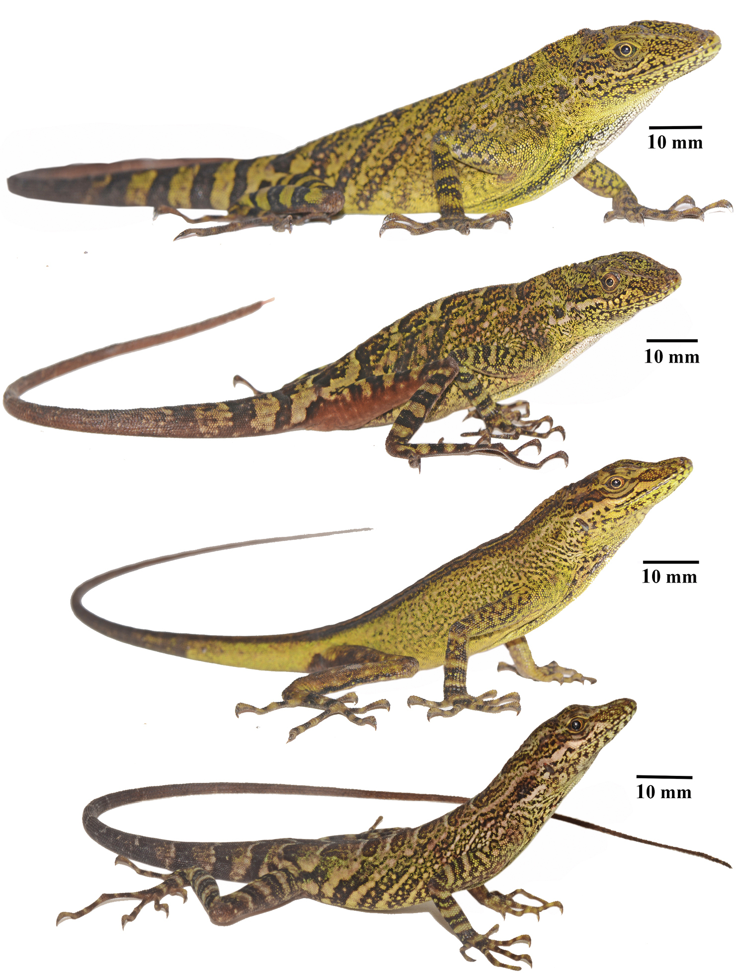
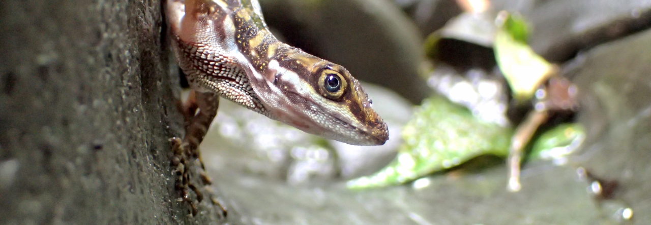
Again, at first glance, it appears to be a green anole. But look at the ear cavity. In this observation of a male A. allisoni, the ear cavity is also more of a gash, instead of a hole. And the large scales on the snout would also imply A. allisoni. What do you think?
Join me on Instagram @dailyanole to follow my adventures!