Category: New Research Page 29 of 67

Bolder Lizards Drop Their Tails More Readily to Compensate for Risky Behavior

(editor’s note: this video was added by the editor. Decide for yourself whether it illustrates the experimental approach described below)

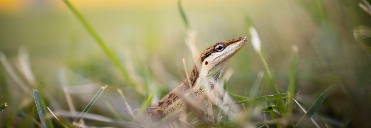
It’s no secret that grabbing a lizard by its tail will often times leave you with the tail rather than the lizard. Why? Because the tail would simply break off. The voluntarily shedding of the tail in lizards (tail autotomy) has fascinated herpetologists ever since the 70s, and it didn’t take long for those people to notice that the propensity for tail autotomy varies extensively among species, conspecific individuals, or even within the same individual at different developmental stages. Four decades have passed, what might be responsible for the variation in tail autotomy is still not entirely clear. In a recent paper, we tried to solve a piece of the puzzle by testing the hypothesis that lizards might autotomize the tail with different propensities to compensate for their intrinsic risk-taking tendency.

Our idea was simple: bolder lizards, due to their behavioral tendency, tend to expose themselves more to higher predation risk. Therefore, selection might favor higher propensities for tail autotomy in bolder lizards as a compensation mechanism. We were also interested in knowing how food availability in the environment might affect tail autotomy. So, we caught a bunch of juvenile brown anoles from the same population in New Orleans and assigned them into two dietary groups: low versus high food availability. After the lizards reached adulthood, we picked out the males and examined the relationship between boldness and the propensity for tail autotomy. (In case you wonder how we measured the propensity for tail autotomy, we refer you to a paper by Stanley Fox, who contributed greatly to our knowledge of tail autotomy.)

And here’s what we found:

The relationship between boldness and the propensity for tail autotomy in the brown anole lizards

Bolder lizards did autotomize their tails more readily as a means to compensate for their risk-prone personality, but only in the group raised with abundant food. Our results helped explain why lizards from the same population autotomized the tail with different propensity. Moreover, our study highlighted the role of food availability in the cost-benefit dynamics of tail autotomy, which has never been explicitly discussed or tested before. Aside from those exciting implications for the study of tail autotomy, our results also have important bearings on broader topics such as the evolution of trait compensation and animal personality. If you are interested in knowing more about this project, check out our recent paper:

CHI-YUN KUO, DUNCAN J. IRSCHICK and SIMON P. LAILVAUX. (2014). Trait compensation between boldness and the propensity for tail autotomy under different food availabilities in similarly aged brown anole lizards. Functional Ecology DOI: 10.1111/1365-2435.12324

Seasonal Shifts in Relative Density of the Lizard Anolis polylepis (Squamata, Dactyloidae) in Forest and Riparian Habitats

displaying on leaf

A. polylepis displaying dewlap.

A commonly observed, but little studied, aspect of tropical herpetology is the seasonal shift in some species’ relative abundance in forested habitat and adjacent, nearby streams. The general pattern is that during the dry season, some species of forest frogs, lizards, and snakes seem easier to detect along streams than in the forest and vice versa during the wet season. Despite this intuitively unsurprising seasonal shift in macrohabitat use being noticed in the 1960s by researchers like Jay Savage and Norm Scott, there has been little work done to document it. In an upcoming issue of the Journal of Herpetology is a paper titled: Seasonal Shifts in Relative Density of the Lizard Anolis polylepis (Squamata, Dactyloidae) in Forest and Riparian Habitats.

The difficulty in documenting seasonal macrohabitat shifts is twofold. First, field sampling must encompass both seasons and be continuous. Second, simultaneous sampling needs to occur in both forest and streams across seasons. For many tropical herpetologists, the opportunity and time for such a study do not come about often. In December 1999, I had this opportunity when I spent three years studying the herpetofauna along the south-central Pacific coast of Costa Rica. I was a young, precocious and budding herpetologist and wanted to understand the ecological habits of all the local amphibians and reptiles. So, out of curiosity I set up transects in a 25-hectare forest patch and a stream that ran through the forest at the Tropical Forestry Initiative (TFI) research station. For 29-months, with the help of field assistants (Deborah Merritt and Yemaya Maurer St. Clair) we sampled the transects regularly, documenting and observing species diversity and habitat use in the forest and stream. While I was organizing the data, an interesting pattern emerged in regard to Anolis polylepis. Of all of the species in the local lizard fauna, A. polylepis showed the strongest seasonal shift in relative density between the two habitats!

Anolis polylepis is the most common anole along the Pacific coast of Costa Rica, reaching densities of up to 300 individuals per hectare (Andrews 1971; Scott 1976). The species can be found in a wide variety of forested habitats ranging from old growth forest to gardens with ample shade trees. In my experience, the only necessary habitat requirement for A. polylepis is shade from a closed canopy. The high density and generalist habits of A. polylepis make it a wonderful study species.

Like many forest anoles, A. polylepis is active in the understory during the day. However, obtaining accurate population counts can be difficult because individuals are wary and can be difficult to detect. For example, A. polylepis will jump to the ground or circle around a tree when observed. This avoidance behavior can be problematic when attempting to obtain reliable counts by increasing the likelihood of missing a lizard. To counter this difficulty, I surveyed for A. polylepis at night, which facilitated easier detection. Anolis polylepis, like many species of anoles, sleeps visibly on leaf tops, twigs, branches and vines from 0.5 to 4 meters above the ground. Thus, it is easier to obtain better counts of relative density for some anole species when lizards are sleeping and inactive. Nocturnal surveys can be very informative for addressing certain questions related to anole biology.

In total, 41 nocturnal surveys were conducted between January 2001 and February 2002, covering one wet and one dry season. We found significant seasonal differences in A. polylepis relative densities between the wet and dry season. During the dry season, A. polylepis density was 0.052 lizards per meter in the stream and 0.010 lizards per meter in the forest. This pattern reversed in the wet season when stream relative density was 0.002 lizards per meter and forest relative density was 0.036 lizards per meter. This seasonal change in relative abundance suggests that wet-dry seasonality influences macrohabitat use in A. polylepis in Costa Rica.

One major limitation of our study was that we did not use mark-recapture. Use of such an approach would give insight into the individual movements associated with our observed patterns. For example, we could test whether lizards are moving large distances to the stream during the dry season, or whether deep forest lizards are moving to moist microhabitats within the forest such as tree buttresses, to name two possibilities.

As with many pilot field projects, ours documents a novel pattern, but raises additional questions. Future work on this issue should extend to other species and regions and use mark-recapture or radio telemetry to elucidate the details of seasonal migrations. An understanding of seasonal movements in environments with distinct wet and dry seasons has implications for how anoles and other herps can tolerate the harsh dry season.

References:

Andrews, R.M. 1971. Food resource utilization in some tropical lizards. Unpubl. PhD diss. University of Kansas, Lawrence.

Scott, N.J. 1976. The abundance and diversity of the herpetofauna of tropical forest litter. Biotropica 8:41-58.

A. polylepis on tree trunk.

Sleeping A. polylepis. Courtesy of Cesar Barrio Amoros.

Sleeping A. polylepis. Courtesy of Cesar Barrio Amoros.


The Genetics of Anolis Lizard Tail Regeneration: (Re)generating Major Internet Buzz

Anolis carolinensis duo with regenerated tails. Photo credit: Joel Robertson.

Anolis carolinensis duo with regenerated tails. Photo credit: Joel Robertson.

Readers of this blog are well aware of autotomy in lizards – self-amputation of the tail – that usually occurs as a result of sub-lethal predation. Readers of this blog are also familiar with the fascinating ability of many lizards to regenerate new tails post-autotomy. Lizards are the closest relatives to humans that can regenerate a fully functional appendage in the adult stage, and understanding the molecular basis of this process can shed light on the latent regenerative capacities in mammals. A new paper published this week in PLOS ONE (Hutchins et al. 2014) provides the first insights into the genetic mechanisms of lizard tail regeneration, using Anolis carolinensis as a model. Via the high-throughput sequencing of RNA from regenerating green anole tails, and the mapping of these sequences to the A. carolinensis genome, the authors describe the genes that are expressed during the regeneration process, shedding light on potential targets for future human therapies.

Disclaimer: I am not an author on the paper, although I do work in the Kusumi Lab with the authors.

While the ability to regenerate a fully functional appendage in the adult phase is likely a deeply homologous trait across animals, it is not uniformly conserved across vertebrates. Fish, as in the zebrafish model (Gemberling et al. 2013), and amphibians, as in the salamander models (Knapp et al. 2013) can regenerate both limbs and tails, suggesting that while the ancestral vertebrate was equipped with this ability, it seems mammals have during their evolution somehow lost it. Evolutionary hypotheses explaining exactly why some taxa lose the ability to regenerate adult appendages are far and wide, ranging from the stochastic to ecologically-specific fitness trade-offs (reviewed in Bely and Nyberg 2010).

But what are the proximate (i.e. genetic) reasons as to why lizards remain strong regenerators while mammals are left holding the short end of the regeneration stick?

ABS 2014: Social Learning in an Australian Skink

Martin Whiting of Macquarie University began his talk at the Animal Behaviour Society 2014 meeting by lamenting how little we know about the social lives of lizards, especially when compared with mammals, certain insects and fish, and most of all, those pesky other reptiles, birds. But the more we examine lizard social behaviour and cognition, the more apparent it becomes that these animals are capable of substantially more complexity than we previously thought possible. Whiting presented some recent research on the Eastern Water Skink, Eulamprus quoyii, that bolsters this view.

Eastern Water Skink, from the Whiting Lab Page

Though not often social, many lizards, including Eastern Water Skinks, live at densities high enough to allow individuals to be within sight of each other. This is a sufficient prerequisite for social learning, defined as learning a task by observing others and modifying one’s own behaviour accordingly. Whiting asked whether Eastern Water Skinks were capable of social learning by training “demonstrater” individuals to perform certain tasks, letting “observer” individuals watch these demonstraters, and then measuring whether this exposure to the demonstraters enhanced the observers’ success at the task at hand.

The answers to Whiting’s questions were not simple. First, age matters—young individuals were twice as likely to demonstrate social learning than old individuals. Second, the task matters—lizards learnt to associate a colour with a food reward by watching others, but the prerequisite task of actually flipping over the coloured cap to access a mealworm was not spurred by observing other individuals do the same.

In the future, Whiting and his students hope to conduct similar experiments with a variety of lizard species that differ in their degree of sociality. These experiments will definitively address the role of learning in shaping the social lives of lizards, and I can’t wait to see they find!

The Fossil Species Anolis electrum Gets an X-ray Makeover

AA readers may remember from previous AA blog posts (here and here) that we have been tackling the field of anole palaeontology; the wonderful world of Amber Encased Anoles. This month, the first paper has been published in the Zoological Journal of the Linnean Society, on the Mexican amber fossil  Anolis electrum (from the collection of UC Museum of Paleontology, Berkeley). And what a fossil!

The amber fossil (left) and x-ray CT reconstruction (right) of one half of the Mexican amber fossil, Anolis electrum.

The amber fossil (left) and x-ray CT reconstruction (right) of one of the two Mexican amber fossils of Anolis electrum. An ant (Azteca sp.) lies behind the right hindfoot. Part of the torso is also preserved (bottom of image). Morphobank images M323739 & M323741.

JMIH 2014: Early Records of Fossil Anolis from the Oligocene and Miocene of Florida, USA

ChovanecKevin Chovanec of East Tennessee Sate University presented one of the most surprising and important posters at the JMIH conference this summer. In his poster, Kevin provides solid fossil evidence for the oldest crown group anole. Working with samples discovered along the Gulf Coast of Florida, Kevin found abundant and well-preserved fossil remnants from anoles. Apparently this material has been around for a while, but has been neglected as attention at these localities focused on identification of mammalian fossils. Kevin has identified the remains of what appear to be at least two species of anoles in deposits that are dated as 26-28 Ma and at least one species in deposits that are 19 Ma. None of this material possesses the traits that are diagnostic for members of the carolinensis series (the only group of extant anoles that was endemic to the United States prior to a wave of recent introductions). His work suggests the existence of a multi-species anole fauna dating back to the Oligocene. A phylogenetic analysis suggests that Kevin’s fossils are members of the anole crown group, but it is not possible to place them with any more phylogenetic precision. He did note, however, that they also lack the transverse vertebral processes that are diagnostic for the β anoles (a.k.a. Norops). The work Kevin presented was part of his masters project at East Tennessee State. I can’t wait to see what other insights emerge from Kevin’s work!


New Anole Distribution Records: Do Lizards in Potted Plants at Home Depot Constitute Range Extensions?

As mentioned in the previous post, the journal Herpetological Review is an excellent resource for anole natural history information. A frequent contribution is range extensions, often by county, for both native and introduced species. Range extensions are important pieces of information for biologists, as accurate county-level distributional data is crucial in many important exercises, such as mapping species richness in a region or identifying range boundaries (and then asking why the range ends in certain areas). This quarter’s issue has the following two range extensions.

Christopher Thawley and Fern Graves report a new county record for Anolis carolinensis in Bullock Co., Alabama, just south of Auburn. This apparently fills a hole in the confirmed range of the species in that part of Alabama.

Cory Adams and friends report an extension of Anolis sagrei range in Angelina Co., Texas. Interestingly, this specimen, as well as a specimen from Nacogdoches, Texas, were found in potted plants in Home Depot and Lowe’s garden departments. The authors posit that these animals turning up in East Texas are not range extensions, as in owing to the expansion of individuals from established ranges, but instead are the result of novel introductions facilitated by interstate transport of goods such as potted plants. If this is the case, these animals could have come from anywhere, not just the invasion front along the Gulf states. In other words, if the potted plants are coming from, say, Florida, then these animals would be leapfrogging their established conspecifics to potentially start new colonies and expand the range.

Adams, CK, D. Saenz, and JD Childress. 2014. Anolis sagrei (Brown Anole). Distribution. Herpetological Review 45: 282.

Thawley, CJ and F. Graves. 2014. Anolis carolinensis (Green Anole). Distribution. Herpteological Review 45: 282.

JMIH 2014: Relative Contribution of Genetic and Ecological Factors to Morphological Differentiation in Island Populations of Anolis sagrei

Wegener

Hanna Wegener, a student with Jason Kolbe at the University of Rhode Island (and an Anole Annals contributor), presented a poster at JMIH on her efforts to identify the factors that drive morphological differentiation among Anolis sagrei populations found on 16 Bahamian islands near Staniel Cay. Hanna investigated morphometric, ecological, genetic, and demographic variation among these populations and, unlike many previous studies, considered variation in both males and females. Although Hanna did find significant morphometric variation among islands and between sexes, she did not find the significant correlation between morphometric variation and habitat use reported in prior work. She also did not find a significant relationship between morphometric and genetic variation.  She did, however, find that population density influences morphometric variation, with lizards living at higher population densities having significantly longer heads than those found on lower density islands. Because these lizards on densely populated islands are also more likely to exhibit evidence of injury from other anoles (e.g., loss of limbs, digits, or claws), it is possible that their longer heads may indicate a response to intra-specific competitive interactions. However,  interpretation of these results remains complicated because there is not a direct connection between injury and intra-specific competition, and the lizards on densely populated islands had longer heads, but not the wider heads that would have been expected if the goal of their morphometric shift was to increase bite force. Hanna undoubtedly has many more exciting questions to investigate with her ongoing research.

JMIH 2014: The Ultrastructure of Spermatid Development within the Anole, Anolis sagrei

Clinger

In a poster at JMIH 2014, Jonathan Clinger of Austin Peay State University found that spermiogenesis (the final step of spermatogenesis during which spermatids develop into mature spermatozoa) in Anolis sagrei is fairly similar to that previously reported in A. carolinensis.

JMIH 2014: Effect of Moisture and Substrate on Egg Water Uptake and Phenotypes of Hatchling Lizards (Anolis sagrei)

Following up on yesterday’s post, more research results from the Warner Lab on egg incubation were presented at JMIH. Corey Cates, a masters student from the Warner Lab, presented his data on developmental plasticity in Anolis sagrei. He used an experimental approach to test whether lizards incubated under dry conditions would survive better in a dry habitat than lizards incubated under moist conditions and vice versa. The idea for the study came from the observation that habitat and substrate differs among small islands in Florida. Some islands are scarcely vegetated and have dry substrate consisting of broken shells. Other islands are more densely vegetated and have dark soil that contains organic matter.

Corey collected 128 breeding pairs from four islands and incubated the eggs using the two different substrates. He also tested two different moisture conditions (wet and dry). He found that lizards incubated under wet conditions hatch on average 4-5 days later and hatchlings were significantly heavier than those incubated under dry conditions. In addition, lizards hatch significantly later when incubated in the soil substrate, which retains moisture longer than the broken shells. Corey further tested whether lizards raised under dry conditions have higher desiccation tolerance than lizards from wet conditions. He measured body mass before and after keeping the lizards in a desiccation chamber. Lizards that had developed under wet conditions lost 5% more mass than lizards developed under dry conditions.

Hatchlings incubated under wet conditions lost significantly more mass than hatchlings incubated under dry conditions.

Hatchlings incubated under wet conditions lost significantly more mass than hatchlings incubated under dry conditions.

This suggests, that plastic responses to different developmental conditions have an effect on physiological traits that might increase survival in a specific habitat. To test this, Corey then released the hatchlings on four experimental islands and measured hatchling survival using a recapture method.

Significantly more hatchlings survived in the open, arid habitat when eggs were incubated under dry conditions.

Significantly more hatchlings survived in the open, arid habitat when eggs were incubated under dry conditions.

He found that significantly more hatchlings survived in open, arid habitats when eggs were incubated under dry conditions. No effect of incubation condition on hatchling survival was found in the shaded, moist habitat.

Page 29 of 67

Powered by WordPress & Theme by Anders Norén