
Anolis sagrei with a regrown tail. Photo by Philip Fortman
Kristin Winchell has the answer. Check it out on her blog, Adaptability. Here’s a shot of the poster she discusses:

Anolis sagrei with a regrown tail. Photo by Philip Fortman
Kristin Winchell has the answer. Check it out on her blog, Adaptability. Here’s a shot of the poster she discusses:
Compared with our extensive knowledge of male-male interactions, we know very little about how females interact with one another. Adding to a growing set of observations, here is some video (taken by my field assistant and seasoned anole videographer Jon Suh) of two bead-tagged female brown anoles mid-battle.
Both females are recent arrivals to this particular tree, and the lizard that remains on the tree at the end is marginally bigger than the one who leaves. Though I don’t think we witnessed the full interaction, I think it’s interesting that the females didn’t use their dewlaps in the course of this fight. This seems to match up with Ellee Cook’s description of a fight between two female A. gundlachi. The use of the dewlap by females has been observed during male-female interactions in A. cristatellus, A. armouri and a few other species, but also during female-female interactions in some Central American anoles. Clearly we’ve got a long way to go before we characterize and understand agonistic encounters and display behaviour in female anoles!
Liam Revell writes:
My co-authors (Luke Mahler, Graham Reynolds, & Graham Slater) and I recently presented a ‘new’ method for placing recently extinct taxa into a backbone molecular phylogeny on the basis of quantitative trait data. I say ‘new’ with quotes, because our methods derives closely, with full credit given where due, from a Maximum Likelihood phylogeny inference approach presented originally by Felsenstein (1981, 2002).
The idea is basically as follows. We start with a time-calibrated molecular phylogeny containing N – 1 species, and a single taxon of interest (the Nth taxon) whose placement in the tree is of interest, but for which molecular characters are missing. If we have quantitative trait data from one or more characters for all N species in the tree, we can use an approach based on Felsenstein (1981) to add this taxon to our base phylogeny using the statistical criterion of Maximum Likelihood. In our article (Revell et al. 2015), we demonstrate that the method works pretty well in theory. In fact, for more than a few quantitative characters & particularly for trees of large size, the method often places the missing taxon in our dataset in a topological position that is identical to its true position. (See figure below, reproduced from our article.) In the figure, white bars show the performance of our method (compared to grey bars which represent placement at random). In all cases, lower values indicate that the estimated tree is closer to the generating tree.
The question you’re probably asking yourself (and quite rightly so) is: what could this possibly have to do with anoles? The answer is that we applied the method to the unusual case of Anolis roosevelti. Anolis roosevelti, as many readers of this blog likely already know, is a mysterious crown-giant anole from Culebra and (probably) the Spanish, U.S., and British Virgin Islands, excluding St. Croix. It is only known from a few specimens and was last collected in 1932. Aside from some unconfirmed reports, it has neither been seen nor heard from since. Unfortunately – and tragically given the impressive nature of this creature – all but the most optimistic anole biologists agree that this species is most likely extinct. (Many of us, the author included, still holds out hope, of course.) The figure below shows the type specimen of this impressive creature. (Figure from our article and image courtesy of the Museum of Comparative Zoology at Harvard.)
 Since no prior investigator has collected molecular characters from this species, and the prospects for so doing in the future are somewhat mixed (for reasons that we explain in the article), we thought Anolis roosevelti would represent an interesting test case for our method. Would A. roosevelti, we asked ourselves, fall out as sister to the Puerto Rican crown-giant, Anolis cuvieri, as sister-to or nested-within the rest of Puerto Rican anoles, or in another part of the tree entirely?
Cabela’s telescopic panfish poles (in both 10′ and 12′ versions) are currently on sale, reduced from $27.99 to $17.49 per pole. These are currently regarded as the best product in the anole noosing game (1,2,3).

Telescopic panfish poles on sale here.
Get them while you can!
Bill Schlesinger, one of the world’s most eminent biogeochemists and President Emeritus of the Cary Institute of Ecosystems Studies, turns out to have an eagle eye for anoles. While on a birding trip to Puerto Rico, his wife, writer Lisa Dellwo, snapped the photos below in the rainforest in the west central part of the island. Which species are they?

Piero Angeli Ruschi from the Ornithology Department in the National Museum in Rio de Janeiro sent the pictures of Anolis punctatus shown here. His story:

It’s raining lizards!
“These are the pictures that I took of one of the Anolis specimens that had fallen that day. They fell from the canopy straight to open ground over an area of ~70 squared meters. About 10 specimens fell within ~3 min. It happened in Santa Lucia Ecological Station, at Santa Teresa, ES, Brazil in late september 2007 during the afternoom while I monitored a woodpecker nest.
The individuals were all the same as the one in that picture…green with a yellow circle around the eye…Those pictures might even include more than one individual—I am not sure if the one photographed on the ground was the same I captured (they stayed knocked out for a minute or so before running from where they landed).

I wish I had more data to publish a communication about it, as well as more evidence to explain the cause of it. My ornithological background leads me to the alternative hypothesis of some sort of “arena,” but I have no idea if such thing can be expected for these lizards.”

Piero Angeli Ruschi in his day job observing birds
At the risk of developing the reputation of being the harbinger of bad news, I’m here to report what seems to be an epidemic of sorts afflicting the green anoles in Gainesville, FL. In the last two years in this town, veteran AA correspondent Thom Sanger and I have noticed a number of very sickly and dead Anolis carolinensis. Here are some photos from last summer:

A sickly green anole that died the next morning. Photo by Thom Sanger.

We saw these animals in the later summer months, and Thom wondered if they might have died from ingesting insects that had been contaminated with insecticides sprayed to control mosquitoes. But a few days ago, my field assistant Jon Suh saw another mysteriously dead green anole, and it’s too early in the year for it to be explained by pesticide. This was in my fieldsite in the UF campus, where I haven’t seen any cats. The lizard also didn’t appear to have any botflies or other large parasites on it (though I’m not sure what that blue spot is…).

It’s worth noting that we have seen no dead brown anoles in the same sites, so it appears that the cause of these lizards’ demise is species specific. Also, we haven’t noticed any dead lizards in the state parks just outside the city, so it seems to be specific to urban areas. Does anyone have any ideas about what might be afflicting these lizards?

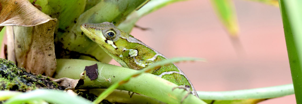
I was just curious if any one could help me identify an anole I found in central Costa rica, near San Ramon in the Alberto Manuel Brenese Biological Reserve. It appears to be a male, and I’m assuming in the Dactyloa genus. My guess would be the Dactyloa casildae, but I am family uncertain. It would be greatly appreciated if someone could help me out! Cheers and good health!

If you’re in Gainesville and come across a site like this, odds are you’re not at a crime scene, but rather Ambika Kamath’s study site, where she’s investigating the social organization of brown anoles. The standard view of anole social structure is that a male brown anole defends a large territory, excluding all other males and courting the several females that reside therein. Ambika’s work to date suggests that reality is a lot more complicated. Ambika provides an in-depth discussion of what she’s doing and why on her blog .


Green anole with a regrowing tail. Photo from Daffodil’s Photo Blog.
Lorenzo Alibardi is conducting detailed studies of what the cells are doing as the tail regenerates. His latest work is now available online in the Journal of Morphology. Here’s the abstract:
Using an antibody against a lizard telomerase-1 component the presence of telomerase has been detected in regenerating lizard tails where numerous cells are proliferating. Immunoblots showed telomerase positive bands at 75–80 kDa in normal tissues and at 50, 75, and 90 kDa in those regenerating. Immunofluorescence and ultrastructural immunolocalization showed telomerase-immunoreactivity in sparCe (few/diluted) mesenchymal cells of the blastema, early regenerating muscles, perichondrium of the cartilaginous tube, ependyma of the spinal cord, and in the regenerating epidermis. Clusters of gold particles were detected in condensing chromosomes of few mesenchymal and epithelial cells in the regenerating tail, but a low to undetectable labeling in interphase cells. Telomeraseimmunoreactivity was intense in the nucleus and sparCe (few/diluted) in the cytoplasm of spermatogonia and spermatocytes and drastically decreased in early spermatids where some nuclear labeling remains. Some intense immunoreactivity was seen in few cells near the basal membrane of intestinal enterocytes or in leukocytes (likely lymphocytes) of the intestine mucosa. In spermatogonia, spermatids and in enterocytes part of the nuclear labeling formed cluster of gold particles in dense areas identified as Cajal Bodies, suggesting that telomerase is a marker for these stem cells. This therefore suggests that all of the sparCe (few/diluted) telomerase positive cells detected in the regenerating tail may represent sparCe (few/diluted) stem cells localized in regenerating tissues where transit amplifying cells are instead preponderant to allow for tail growth. This observation supports previous studies indicating that few stem cells are present in the stump after tail amputation and give rise to transit amplifying cells for tail regeneration.
Powered by WordPress & Theme by Anders Norén

