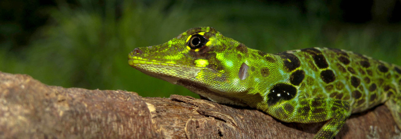

A mourning gecko (Lepidodactylus lugubris) climbing vertically on glass with the help of its impressive toe pads.
I think most people visiting Anole Annals could argue that the adhesive digits of anoles are some of the most fascinating aspects of their biology (or maybe I’m just biased). Digital adhesion is accomplished through toe pads: a collection a broad, modified plantar scales which bear thousands upon thousands of microscopic, hair-like structures (i.e. setae). Through frictional and van der Waals forces, these collections of setae allow toe pad-bearing lizards to easily access vertical surfaces and exploit habitats many lizards cannot. Shockingly, adhesive toe pads have independently evolved several times across lizard evolutionary history (at least 16 times by recent estimates) — once in the common ancestor of anoles, once in a clade of southeast Asian skinks, and 14 times in geckos. Both within and between the different evolutionary origins of toe pads, there is substantial variation in toe pad size, shape, number of scansors/lamellae, and position of the adhesive apparatus.
In our recent study, my collaborators and I took the first steps to characterize how embryonic development is modified to achieve this incredible diversity. Using embryonic material my coauthor Thom Sanger collected as a postdoctoral researcher in Marty Cohn’s lab, in addition to embryonic material I collected over the course of my Ph.D. training in Tony Gamble‘s lab, we aimed to compare embryonic digit development of ancestrally non-padded lizards with that of anoles and padded geckos. We used a model clade approach to broadly sample anoles and geckos, although some species breed more easily in the lab and have more embryological resources than others. All together, we sampled a range of toe pad morphologies in both clades (trunk-ground and trunk-crown Anolis ecomorphs and leaf-toed and basal pads in geckos). To help polarize the developmental changes leading to the origin of toe pads, we also included two ancestrally padless species in our comparisons. After the collection of these diverse embryos, we used scanning electron microscopy (SEM) to characterize scale morphology of the digits throughout embryonic development.

By comparing embryonic material of anoles and geckos, we essentially span the diversity of squamates in a single comparison.
Because of the ~200 million year divergence between anoles and geckos and dramatic differences in adult morphology, we anticipated that we would see stark differences in the developmental origins of toe pads in these species. To our surprise, we found striking similarities in toe pad development between all of the pad-bearing species we examined. We found that toe pads develop after digit webbing recesses. In all pad-bearing species, ridges that become the adhesive scansors and lamellae first form in the distal half of the digit. Throughout development, new ridges begin forming in the proximal direction while the previous ridges begin to grow laterally. Elaborations and derivations in toe pad form, such as bifurcation, occur in the latter-half of embryonic development. The presumably ancestral pattern of plantar scale development we observed in our leopard gecko and fence lizard embryos (both species lacking adhesive digits) demonstrated that scale ridges form all at once along the length of the digit. These differences are similar to those documented between developing non-padded gecko tails and padded tails of crested geckos. This means that anoles and geckos have converged on a similar developmental process! We suggest that toe pads are initially formed through a major repatterning of digital development and then variation is achieved through relatively minor “tinkering,” through either timing or location of developmental patterns.

Scanning electron micrographs (SEMs) of embryonic lizard digit development, progressing from early development (left) to late development (right). The pad-bearing brown anole (Anolis sagrei) and mourning gecko (Lepidodactylus lugubris) have converged on scansor ridges forming in a distal-to-proximal direction, while the paddles leopard gecko (Eublepharis macularius) has scale rows forming all at once along the length of the digit. Lizard photos courtesy of Dr. Stuart Nielsen.
This is by no means the end of this story. We’ve just scratched the surface and there are a several directions to head in. A logical next step is to characterize histological organization through toe pad development. From there, characterizing the genes involved in toe pad morphogenesis, in tandem with the possibilities of new gene editing technologies, would allow us to test mechanisms of toe pad formation and how variation is generated. And, of course, characterizing toe pad development in other species (such as the secondarily padless Anolis onca) may elucidate further conservation or derivation from the trends we found. This is an exciting time to be a toe pad biologist!
- Making the Fancy Feet of Anoles and Geckos - February 1, 2022
Leave a Reply