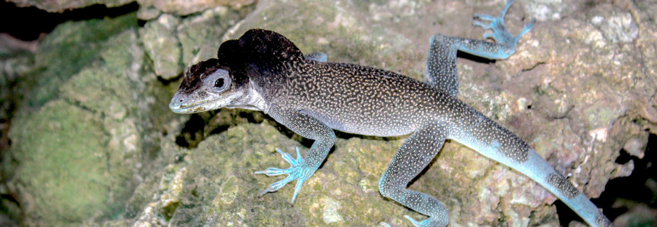

Melissa Kemp with a Puerto Rican crown-giant anole (Anolis cuvieri). Photo by Melissa Kemp
Paleobiologist Melissa Kemp spends a lot of time overturning assumptions. Her excavations don’t involve digging bleached bones out of windswept deserts, but looking for partially preserved lizard fossils in dark, dank jungle caves. Last month, she published a study tracking human-driven species introduction in the Caribbean through the region’s 7,000 years of human habitation—challenging the idea that “restoring” Caribbean biodiversity means taking it back to where it was before Christopher Columbus arrived in the so-called New World around 530 years ago.
Kemp, who runs a lab and teaches integrative biology at the University of Texas at Austin, opened up on Twitter last week about her experience as a Black scientist and outdoorswoman, under the hashtag #BlackInNature—as part of continuing conversations about race in America following the killings of Ahmaud Arbery, Breonna Taylor, and George Floyd, and the harassment of Black birder Christian Cooper. She spoke with NOVA about decolonizing environmental restoration, tropical fossil quirks, and the joys of time outside.
Alissa Greenberg: Let’s start with the hashtag #BlackInNature, which you’ve used in tweeting about your love of spending time outside. What’s important about that time in nature for you? What do you feel like it does for you physically and emotionally?
Melissa Kemp: I love spending time in nature. I live in Austin now, in a much more urban environment than I was raised in. But it’s still very rejuvenating just to go outside and look at the sky, look at the plants, find animals on the property and just see that there’s life there. Even when I’m doing my field research, there’s bursts of active work where we’re hiking through the rainforest trying to get to our site. But then when we get there, it can be very slow. The work that we’re doing is very meditative. So nature is very therapeutic for me. It’s played a very important role for me personally and professionally.
Particularly thinking about the COVID crisis, so many people are trying to find solace in nature during this time. And I think, now more than ever, it really needs to be accessible to everyone, with everything that’s going on—not only to make discoveries in and learn about, but just to enjoy and to feel comfortable enjoying it.

AG: You mentioned in a tweet that you grew up exploring outside on land your great-great-great-grandfather bought after emancipation. So your family has been there ever since?
MK: Yes. I grew up in Maryland, outside of Baltimore. Not really that far from any city, but very, very rural and situated near a state park. And because of that, I had a lot of nature at my disposal. I grew up listening to stories. My mom and my aunts and uncles would tell stories about how they would go out in the woods and explore. So I always had a connection to nature. I never questioned that connection because I felt like I lived in it—even just knowing that my family had been there for so long. The church cemetery was across the road, so I could go in the woods and see the graves of my ancestors.
AG: You also mentioned in that same tweet that your grandmother taught you to mark recapture, the biology technique to help estimate animal populations.
MK: Especially during the summers when my cousins would be there, and my grandmother had all these kids to deal with, we would go around looking for animals. We always found turtles, eastern box turtles. We would write our initials on them in nail polish, which we really probably shouldn’t have been doing, and take care of them for a night, then release them. She would always tell us, “Look for your turtles,” and we would find them again. Sometimes years later, we would find a turtle and be like, “Wait, that’s MK, that’s my turtle!” She really encouraged us to just go out there and explore. I think it really rubbed off on me.

AG: How did you end up working in integrative biology? And why did you choose to focus on islands?
MK: I didn’t come into science in the most traditional way. When I was growing up, I always thought I was going to be an artist. I went to art magnet schools as a kid and trained at a really high level, mostly painting and drawing. I still approach science in a way that is similar to how I approach art. This diligence of working on something for a very long time and also being open to feedback from others to make the work better. Art is a very iterative process. It can take years before a piece is done, and it’s not a sprint, it’s a marathon. I think a lot of the same concepts apply to science as well. And my eye for detail maybe helps me find nuances in materials I look at. When I’m working with fossils I’m constantly looking at different shapes and looking for differences in structures of the different fossils I’m handling.
I study how biodiversity in tropical regions originates both through processes of extinction and diversification, as well as colonization. Particularly, I’m interested in how changes in the environment then impact the different communities of organisms that we have. I’m interested in these past instances of change that we can see through the fossil record, because it’s the key to really understanding the biodiversity that we have today, but also helps us understand how biodiversity might change in the future.
Islands in particular are really interesting biologically, and there’s been a wealth of study of diverse life-forms of islands, particularly lizards. But we don’t really have as much literature on fossil occurrences of lizards. That’s why I started focusing on islands, because we really don’t have a thorough understanding of how we got to present day biodiversity.
Human-driven biodiversity change in the Caribbean did not start in 1492. There is a 7,000-year legacy of change.
AG: You recently published a paper that delves pretty deeply into that topic. Can you summarize what you were looking for and what you found?
MK: We were really interested in investigating how humans have modified the biota of the Caribbean, particularly through which species they’ve introduced. This paper is really thinking about what we are adding to the islands—and what are the impacts of what we’re adding? How do those introductions scale across time? We developed a database of species introductions by going through the archaeological literature, as well as the paleontological literature, to get a sense of what we know about species introductions, and then also what we don’t know.
I think one of the biggest takeaways is the fact that humans have been engineering the landscape for millennia, particularly these places that we think of as recently perturbed. Human-driven biodiversity change in the Caribbean did not start in 1492. There is a 7,000-year legacy of change. We have these terms that I don’t think are representative of the biodiversity of the Caribbean, like “New World.” They’re really terms steeped in European colonization, referring back to the Caribbean as something being “new” to Europeans, when people had been living there for a very, very long time.
When we think about what the Caribbean used to look like, if you ask somebody on the street that question, they’re probably going to think about before Columbus and Europeans came. And we are definitely interested in that period, very much so. But we’re also interested in what it looked like before any humans arrived. And we really want to acknowledge how Indigenous groups in the Caribbean moved around, what species they were bringing, how they were using species, and how their manipulation of the landscape changed biodiversity in the Caribbean.
AG: Why is it important to ask those questions? Not just culturally but also scientifically?
MK: We have to be realistic about what’s actually feasible. The ideal restoration target probably in the head of a lot of people in the public would be what the environment was like before we came and messed it all up. But it’s not a realistic target, in part because we’ve lost so many species that were in the landscape before humans arrived.
What we see in a lot of systems after an extinction of one animal is that you have an extinction cascade where other things go extinct because organisms are interconnected with one another. They don’t exist in silos in the landscape. So, if we remove a pollinator, the plants that were pollinated by it might also undergo decline. They might also go extinct. And that might affect soil erosion, for example. Maybe their roots provided important structure for soil. And if you’re eroding soil, maybe you’re interrupting something else’s habitat. So we would want to restore this system so that that plant is there, and so that that plant is pollinated by an organism in the system—but it might not be possible to do all of those things. So, we have to think about, what is it that we are hoping to accomplish through restoration?

Maybe another takeaway from our research would be thinking about introductions of species and what they mean biologically. There’s a lot of different terminologies that we use for introduced species—I think one of the most common one is “invasive,” because we often think about the negative impacts of species introductions. Certainly there were instances where species had very, very negative impacts, but then there are also instances where species don’t seem to be having a negative impact on the environment and maybe are actually doing good things for those ecosystems.
The Caribbean had a lot of endemic mammals prior to human colonization. There were monkeys and a lot of really unique mammals—like these animals called Nesophontes, which are these shrewlike insectivores that are no longer there. We think that a lot of them were really important pollinators in the ecosystem that were then lost. But with the introduction of new birds to the Caribbean, for example, it’s possible that some of that loss of pollinators, that ecological service, may have been restored.
AG: I’ve read that paleontology in the tropics is particularly difficult. What makes it so hard?
MK: The environment of the tropics is not really conducive to fossilization processes. You need stable temperatures, ideally, cold temperatures and dry weather, to get good fossilization—and things covered up really quickly. The tropics are very hot and very humid. It’s just so hot, it’s so wet, that it’s going to erode away very quickly compared to something that’s in the Arctic, for example. There’s much more rapid disintegration when it’s hot and humid. Microbes breaking things down is certainly part of it. Also exposure to UV light—there’s physical damage being done to the material as well as biological damage.
So we don’t have a lot of fossilization in the tropics, but we do have some, particularly in environments where the material is somewhat shielded. Almost all of the work that I conduct is done in caves. We’re going through often heavily forested areas, in limestone landscapes where the humidity and precipitation cuts through the limestone and creates cavities. Usually materials get in there through water flow—when, let’s say, there’s a hurricane.
A lot of it is very fragmented bones. We’re not getting a full lizard, with the skin removed and the bones in perfect position. Some of them have features that are identifiable, some of them do not. I think that’s one of the reasons people have been turned off studying them in the past. A lot you can look at with the naked eye and figure out what part of the skeleton it is, but some of them you need to look at under a microscope.

AG: So what techniques have you used to sort of get around those challenges?
MK: The biggest thing is just not to disregard the data that do exist and brush it aside. If you want to get material from the tropics, it’s not necessarily going to be very, very flashy in the same way that maybe a T. rex skull would be, but there are valuable data out there. Another thing that we do to get around some of the challenges is we just keep on looking. It requires us to interface with people in that area, talking to people about where caves are, if they’ve ever seen fossils.
So it’s very much a community effort, in terms of the work that we do. Finding sites with the help of local people. So local people who are out there exploring the caves for fun often have been a real godsend for us, very helpful in orienting us on the land. They’re almost always happy to show us and then interact with us when we tell them more about what we’re doing. And that’s always really fun.
I think it’s a function of where I do fieldwork that I have always felt safe in the field. I work in spaces where there are very diverse cultures that are not my own, often cultures where Black people are dominant or brown people are dominant. When I worked in Guadeloupe, for example—a French-speaking island in the Lesser Antilles where the majority of people are Black and Creole—if I kept my mouth shut, people just assumed I was from Guadeloupe. Being an outsider hasn’t been a source of fear in those landscapes.
Just seeing my grandmother as a Black property owner in a small town where there were not many other Black property owners was very inspiring. I don’t know if it’s the right word for it, but I felt that the outdoors belonged to me and that it was my right to be there.
AG: To that end, what’s important about the conversations we’re having now, around the incident with Christian Cooper and the #BlackInNature and #BlackBirdersWeek hashtags? What would you hope might come out of them?
MK: It’s important because it reinforces, particularly for us as Black people, that we belong here, that this country is ours. We had a very, very instrumental part in creating what we have today in this country, even as we continue to be oppressed. I think it’s also important for non-Black people to hear that as well, that they recognize those contributions. I feel very privileged to have had a very positive association with the outdoors all my life. Just seeing my grandmother as a Black property owner in a small town where there were not many other Black property owners was very inspiring. I don’t know if it’s the right word for it, but I felt that the outdoors belonged to me and that it was my right to be there.
I heard messages going through school from other people like, “Oh, nature is more of a white space.” But I really rejected those messages because I had this pride in my family history, and my connection to the land, and my family’s connection to the land. I just felt like everyone else had it all wrong, that they just didn’t know the history of this country well enough—how tied, for better or for worse, African Americans are to the land. It’s a very painful history, sometimes, to think about how many Black Americans got to this country, why we were brought to this country in the first place, to work the land that white people didn’t want to work.
For me, at least, learning that history has really made me feel more grounded in the space that I occupy. I’ve always felt grounded because of my very strong family history. But I know a lot of Black Americans don’t have that. Talking to distant cousins that I share lineage with further back, helping them learn about the history of enslavement of our family, has really helped ground them as well.
Whenever people may have made snide comments—“I’m afraid of the woods,” or, “The woods is a white space”—I’m very comfortable with my story and being like, “Well, I’m here. My family’s here. They’ve been on this land for six generations. You can’t tell me it doesn’t belong to me.”


