
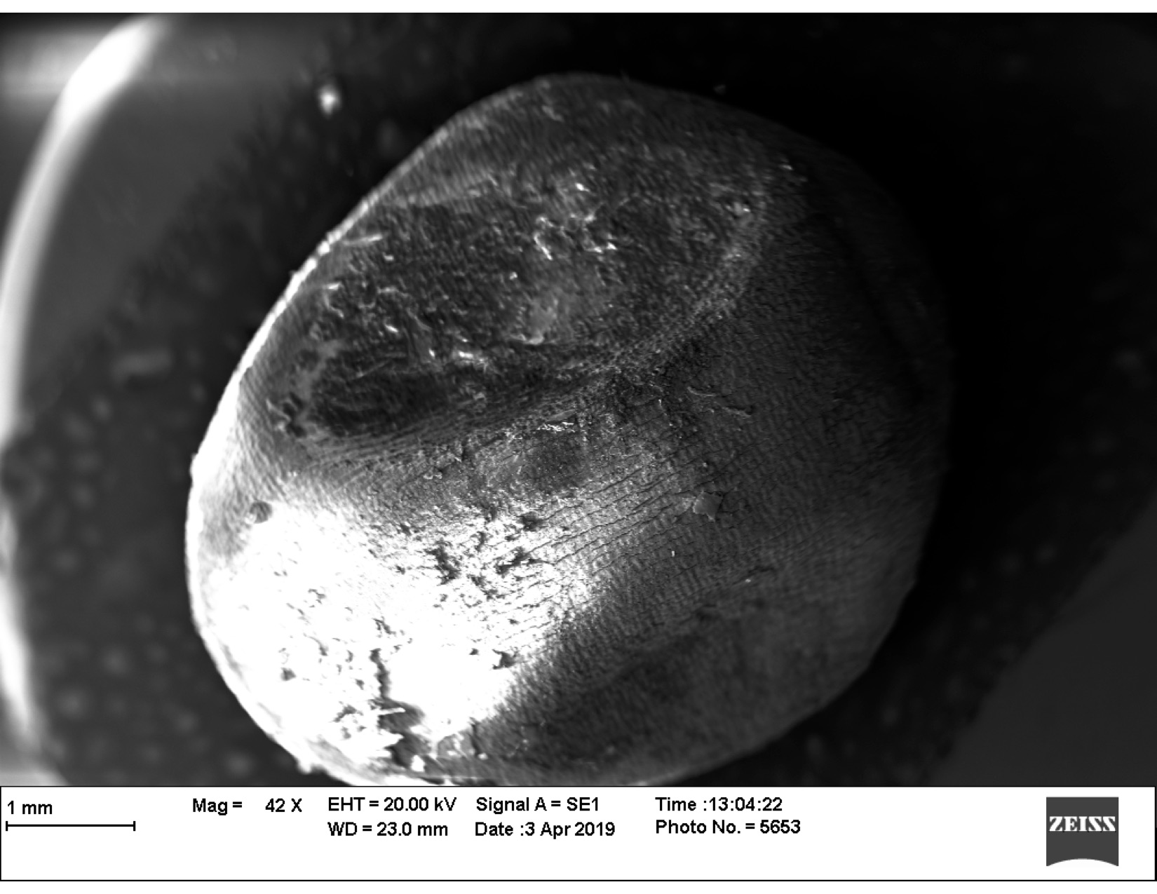
Scanning electron microscopy (SEM) is a technique that utilizes electron beams that interact with and reflect the surface of a viewed specimen. These reflections allow the evaluation of surface topology and ultrastructure and give high-resolution detail about external structures and cellular arrangements (Goldstein et al. 2017). To create a reflection on specimen surfaces, a thin layer of gold is mechanically applied through a process known as “sputter-coating.” Recently, graduate students at Auburn University had the opportunity to view their own collected biological samples with SEM through an Applied and Environmental Microbiology course taught by Dr. Mark Liles.
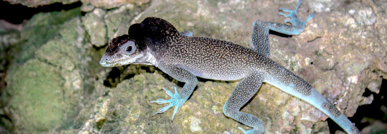
As a student in this class, I had the opportunity to view a chosen sample under this process. While I highly debated bringing in an anole fecal sample (which would have been gold-coated and placed on my desk for a lifetime), I decided to view a recently dried, fertile A. sagrei egg collected from the lab of my advisor, Dr. Daniel Warner. The microbial communities on the surface of this egg were most likely highly impacted by the influence of drying (see image descriptions below); this is due to sample preparation required by conventional SEM, whereby water vaporization will distort images if the sample is not completely dry. Part of my research within the Warner lab involves investigating the microbial communities on the external surface of eggshells; thus, this class has provided an excellent opportunity to explore how varying environmental factors can influence eggshell microbiomes. The photos taken and attached were observed on 03 April 2019.
In Image 1 at 42X magnification, you can see the influence of drying from the large indentions on the egg as well as horizontal cracking within the surface itself. However, under closer inspection fungal and bacterial structures begin to appear. In Image 2 at 397X magnification, you can view a filamentous structure that we predict to be fungi. One of the limitations of SEM is that while structures can be easily viewed, they may not always be as easily identifiable. At 1,500X and 1,5700X, we can see a magnified image of a fungal root (Image 3) and potential bacterial cells above the spiral filamentous structure (Image 4).
 Image 2. SEM image of A. sagrei egg at 397X magnification.
 Image 3. SEM image of A. sagrei egg at 1,500X magnification.
 Image 4. SEM image of A. sagrei egg at 1,5700X magnification.
The images above highlight the interesting use of SEM for reptilian eggs, especially those small enough to be entirely encompassed under a microscope (< 1.5 mm long). SEM observations can also be used to elucidate differences in eggshell structures, thickness, and porosity (Heulin et al. 2002). Additionally, SEM use within the classroom setting has allowed students to gain applicable skills and techniques, as well as their own photographs (Beane 2004).
References:
Beane, Rachel J. 2004. “Using the Scanning Electron Microscope for Discovery Based Learning in Undergraduate Courses.” Journal of Geoscience Education 52 (3): 250–53. https://doi.org/10.5408/1089-9995-52.3.250.
Goldstein, Joseph I., Dale E. Newbury, Joseph R. Michael, Nicholas W. M. Ritchie, John Henry J. Scott, and David C. Joy. 2017. Scanning Electron Microscopy and X-Ray Microanalysis. Springer.
Heulin, Benoit, Samuele Ghielmi, Nusa Vogrin, Yann Surget‐Groba, and Claude Pierre Guillaume. 2002. “Variation in Eggshell Characteristics and in Intrauterine Egg Retention between Two Oviparous Clades of the Lizard Lacerta Vivipara: Insight into the Oviparity–Viviparity Continuum in Squamates.” Journal of Morphology 252 (3): 255–62. https://doi.org/10.1002/jmor.1103.
- SICB 2022: Anole Nesting Behavior under Predator-Presence! - January 15, 2022
- SICB 2022: Let’s Chat about Lizard Sperm! - January 13, 2022
- SICB 2022: Lizards and Lead: What’s Going on with Anoles in New Orleans? - January 11, 2022
Leave a Reply