Yes, it’s true. A “third eye” does exist, not only in the ancient Hindu literature and the new age imagination, but in birds, amphibians, reptiles, fish, lampreys, and hagfishes. We’re talking about the pineal gland, a small organ located on top of the brain, just underneath the surface of the skull. Although it doesn’t have visual capabilities in the image-forming sense, it is intrinsically photosensitive, responding to light signals without any help from the lateral eyes. (Mammals, including humans, have a pineal gland too…but it has lost the ability to detect light).

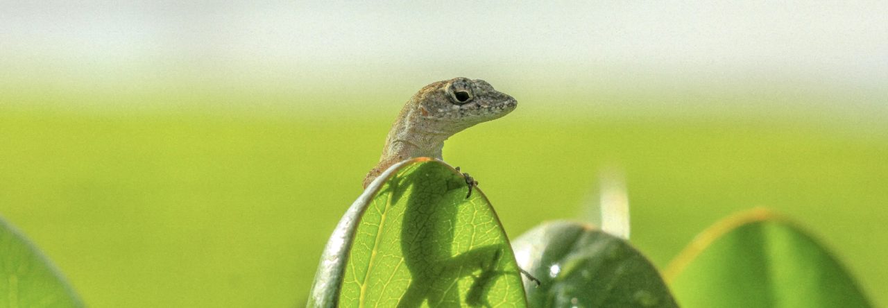
You can see the parietal eye on top of this anole’s head (it’s the tiny circle in the middle). The pineal gland can’t be seen externally, but it’s just posterior to the parietal eye and right underneath the surface of the skull. Photo credit: TheAlphaWolf, License:Creative Commons Attribution-Share Alike 3.0 Unported
Anoles, and some other lizards, actually have two “third eyes,” one being the pineal gland, and the other being the parietal eye, which can be seen in the picture above. Both the pineal gland and the parietal eye are involved in the detection of irradiance (the brightness or energy of light) and temperature, and the two structures are connected neurally. The pineal is located just ventral and posterior to the parietal eye, and cannot be seen from the outside. This little organ functions as a photo-thermo-neuroendocrine transducer, which means it translates light and temperature information into a hormonal signal the brain and body can understand. The hormone produced is melatonin, which is typically synthesized at night and indicates the presence of nighttime to the animal. (Betchya thought the eyes did that! They do, in mammals. But even in mammals, the eyes send some information to the pineal gland to control melatonin production). In virtually all animals studied to date, melatonin is rapidly suppressed upon exposure to light. Surprisingly, in a study by Herbert Underwood, it was found that light does not suppress melatonin levels in A. carolinensis. This was extremely surprising since, in anoles, melatonin is one of the main signals controlling arousal and activity and, as we are all aware, anoles are strongly diurnal. Moreover, the Anolis pineal is photosensitive and contains a circadian oscillator that produces melatonin rhythms. This circadian rhythm is synchronized by light-dark cycles.
What’s going on? If melatonin is important for controlling the timing of activity, shouldn’t the levels of this hormone correlate with the presence of light and darkness?
In a recent paper published in Comparative Biochemistry and Physiology (and supported by NSF Grant #0910075), University of Virginia graduate student Ashli Moore decided to study the photic control of pineal melatonin in other Anolis species, thinking that perhaps A. carolinensis was strange, or that photosensitivity of the pineal varied across clades or according to photic habitat. She measured melatonin rhythms under normal light-dark cycles and under cycles interrupted by a two-hour nighttime exposure to light, expecting to see suppression of nocturnal melatonin during light exposure. You can read all the gory details here. The punchline is that suppression of melatonin does happen in anoles, but it is very, very weak compared to other vertebrates. Moreover, the effect varies among species. It is weakest in A. carolinensis and A. sagrei, and strongest in Puerto Rican species A. cristatellus, A. gundlachi, and A. evermanni. There didn’t appear to be any correlation between photic habitat and photic suppression of pineal melatonin, although one might need to test additional species to detect such a correlation if it exists. So for now, the reason for the species variation is unknown, but might have something to do with a latitudinal gradient. More interestingly, perhaps, is that pineal function in anoles is unexpectedly unique. This study raises many more questions than it answers, so stay tuned for more work on the Anolis pineal from Moore and colleagues!

Figure 2 from Moore & Menaker showing melatonin profiles in five species of anoles. Controls group means are shown with black boxes. Light pulse groups are shown in white boxes. The light pulse was given during the second night. Suppression of melatonin by light was relatively weak and varied among species.
- Sensory Ecology of the Third Eye - June 14, 2012
- Anole Visual Ecology, Sans Vision - January 13, 2012
- Surprises from the Anolis “Third Eye” - September 1, 2011
Bora Zivkovic
Very interesting. As a former Underwood student (though I did not work on Anolis, I did the quail work) I like to see this line of research continued. Unfortunately, I cannot access the paper itself – is there a way I can get a copy by e-mail? Also, I found it curious that you refer to yourself in the third person in this post 😉
ashlimoore
Hi Bora, sent you an email with the pdf. Glad someone would like to see this line of research continued! The authors thank you for your interest 🙂
Bora Zivkovic
Thank you. And say Hi to (my academic grandfather) Mike.