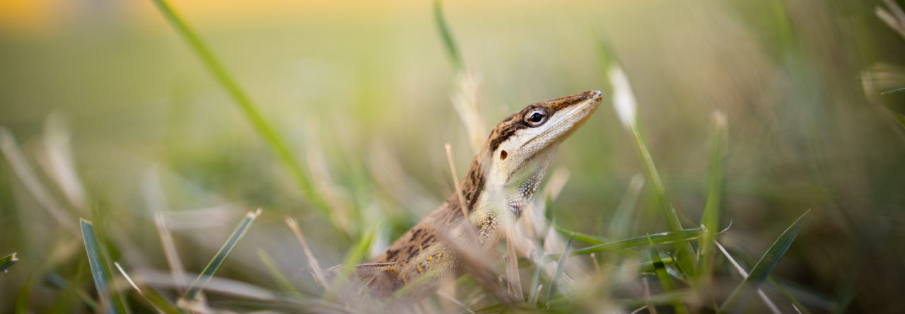

While recently working with scanning electron microscopy, I came across something interesting and I need some help identifying it. As a graduate student in Dr. Dan Warner’s lab at The University of Alabama at Birmingham, I am finishing up my thesis work on the adaptive significance of phenotypic plasticity in Anolis sagrei caused by incubation moisture and substrate. I have found significant differences in the level of desiccation tolerance in individuals incubated under wet and dry conditions. In pursuit of the mechanism by which these differences occur, I was using SEM to observe the spacing of the scales of individuals within my treatment groups. In doing so, I came across a toe of one individual and found several structures which I could not identify. The structures (circled in the picture above) are located on the scales around the toe pad and circling around the toenail. They appear to be hair-like projections.
Despite continued searching and communications with several others, I still cannot identify these structures. Any help or direction would be greatly appreciated.
- Identification Needed: Unknown Structures Found on Anolis sagrei Toes - January 31, 2015
Kenneth Barnett
COOL. Wild guess: The hairs could be a part of mechanoreceptors?
Kevin de Queiroz
Those are called scale organs. There is a fairly extensive literature on them in squamatan reptiles, most of which is from a systematic perspective (they vary in morphology, number per scale, and distribution on the body) rather than a functional one, though they are thought to be mechanoreceptors.
Thomas Sanger
I remember seeing some of these structures in Ernest Williams’ collection of SEMs at the Harvard MCZ. I don’t remember what they are off hand (or how well the figures were labeled), but that might be a place to start if all else fails. You may also want to bump up the magnification on the SEM just to make sure that you are properly identifying them. These structures fill the field of view in the Williams SEMs that I am remembering.
Kurt Schwenk
Kevin is correct. Scale organs are pretty common among squamates. Not all of them have the hair-like structure. N.B. Ananjeva did quite a lot of work on skin ultrastructure. Here’s one of her papers that notes the presence of scale organs on the labial scales of A. equestris: “The Skin Sense Organs of Some Iguanian Lizards.” N. B. Ananjeva, M. E. Dilmuchamedov, T. N. Matveyeva. Journal of Herpetology 25(2 ):186-199. As Tom noted, Ernest Williams did some work on scale ultrastructure in collaboration with Jane Peterson. I know some of that was also published in J. Herp. Also look for German literature by M. von During (umlaut on u) and U. Hiller.
Lars
Check this reference
Paul Klawinski
I have seen these on the dorsal surface of gekkonids (Hemidactylus turcicus) complete with the hair. They were curled down toward the distally adjacent scale on the toes and I assumed they were scale organs involved in fine-scale proprioception as geckos have similar requirments (as anoles) for fine-scale toe movement as they coordinate movement of the toe lamellae.