 Jumping on the 3D bandwagon that has infested Hollywood, I wanted to introduce the Anole Annals community to the newest tool being employed to study Anole diversity and evolution, High Resolution X-ray Computed Tomography, or CT scanning for short.
HRXCT is a tool that uses x-rays to visualize the internal geometries of opaque objects. It is similar to the CAT scan you would get at a hospital, but with high-power x-rays so higher resolution. It is perfect for museum specimens because it is non-destructive; you can study skeletal morphology without removing skin or flesh, unlike the skeletonizing or clearing and staining methods as previously described here. In this first blog post on HRXCT of anoles, I shall explain how the scan process works and how the data are collected.
First, the specimens are prepared for scanning. I have developed an approach whereby I scan several lizards of the same size in one go. This maximises my scanning productivity, since in a 1 hour scan, I get 6 lizard skeletons. Scan resolution is measured in voxels (volume pixels), and is determined by the size of the object in the 2000 x 2000 pixel detector panel. The resolution is the same for 1 lizard as it is for 6, as long as they are all of similar size. for 45mm snout-vent length lizards, I get about 0.045mm resolution. Below is an overview of the sample mounting; the alcohol-preserved anoles are wrapped in alcohol-soaked cheese cloth, sealed in plastic bags, and mounted in oasis foam.
Inside the scanner, 3142 x-ray projections are taken as the specimen rotates 360 degrees. The computer receiving the projections then starts the reconstruction; projections are radially aligned and reconstructed into a volume.

Then using software for editing CT volumes, I segment each lizard from the volume and save it as an image stack. On the slices above, you will see different the different materials are represented by different gray values – black is air, grey is soft tissue, white is bone. I segment the skeleton by thresholding the slices and keeping only the “white” voxels. This results in a rendered skeleton.

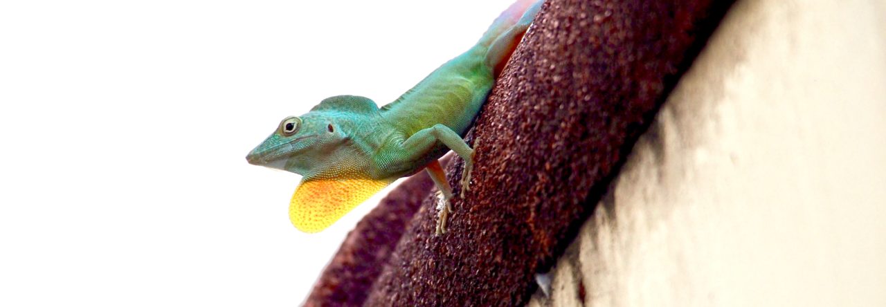
These 3D skeletons are very detailed and reveal the complex geometry of skeletal variation in Anolis. Watch this space for some of the cool things we find using CT, and how the CT scans will aid our research on anoles. And bonus points to anyone you can name the species pictured here!
- 20-Million-Year-Old Fossils Reveal Ecomorph Diversity in Hispaniola - July 27, 2015
- Anolis proboscis: Ugly and Famous - September 5, 2014
- The Fossil Species Anolis electrum Gets an X-ray Makeover - August 14, 2014
Yoel Stuart
Anolis longiceps!
Em Sherratt
Haha, tricked you, since the one pictured live isn’t the skeleton. Keep guessing!
Rich Glor
I’m going to assume that this is a Greater Antillean species (just a hunch). If this is the case, it must be a trunk-ground species. The head is too robust to be a member of the sagrei group and doesn’t appear long enough to be the Jamaican species (A. lineatopus). The skull also looks a bit wider than I’d expect in a Puerto Rican trunk-ground anole. My guess, therefore, is a cybotoid trunk-ground anole from Hispaniola. If we need to guess a species I’m going to go with the most common: Anolis cybotes.
Em Sherratt
Rich, you are very close. It is in fact the holotype of A. marcanoi (MCZ R131876). A fine male specimen. CT scanning will potentially be very beneficial for museum research of anoles – providing digital records of the types, which can be made available to anyone who cannot visit the musuem.
Rich Glor
Do I not get the bonus points? Given that its hard to tell cybotes from marcanoi even with their skins on, I think I got as close as possible from a CT…
Em Sherratt
Ok Rich, You can get the prestige of guessing correctly ;).
Incidentally, yes Talia, the detail is beautiful. It comes down partly to the fact that the specimen was quite large (55mm SVL) and so fully ossified, it is still well preserved (the bone has not deteriorated as can happen with long-preserved alcohol specimens), and the overall resolution of that scan was 0.0556 mm. It has been more tricky with the juveniles (that have more cartilage) and those old specimens that had perhaps been left in formalin for too long – those are still pretty but not up to my perfectionist standard!
Talia
Wow, I can’t believe the amazing resolution you can get on those scans!
marthamunoz
Question for you, Emma. Does those images get uploaded to Digimorph, or to the MCZ database? Where do you put them so that they’re freely accessible? Beautiful images, by the way!
Em Sherratt
Martha, dorsal, lateral and ventral views of the rendered skeleton are being added to the MCZ Herpetology Database (http://www.mcz.harvard.edu/collections/searchcollections.html) as part of the specimen’s file, just as photos of all MCZ Herp types. I have done about 100 specimens from the Dominican Republic like this. My plan is also to make interactive 3D skeleton models. Watch this space!