Author: Thomas Sanger Page 3 of 6

Thom Sanger is an Assistant Professor at Loyola University in Chicago. His lab specializes on understanding the developmental bases of Anolis lizard diversity.

Distribution of Keratins in Lizard Scales

Keratins are the structural proteins of skin, hair, nails, feathers, and scales. There are 54 described  keratins in humans, a subset of which has also been found in the green anole genome. Distinct combinations of keratins in skin appendages are what give these tissues their unique properties such as flexibility, rigidity, or cornification (i.e., the process of forming an epithelial barrier). Lizards have a number of specialized scale types, likely due to the distinct distribution of keratins in those scales. Dating back at least a decade, Lorenzo Alibardi and colleagues have been making great progress describing the keratin gene family in lizards and describing the distribution of these proteins across the body. Alibardi has recently added to this long series with a description of keratin localization in the lamellae of Anolis carolinensis. Because I am not an expert in keratin biology I will let the Abstract give you the details:

ABSTRACT Knowledge of beta-protein (beta-keratin) sequences in Anolis carolinensis facilitates the localization of specific sites in the skin of this lizard. The epidermal distribution of two new beta-proteins (betakeratins), HgGC8 and HgG13, has been analyzed by Western blotting, light and ultrastructural immunocytochemistry. HgGC8 includes 16 kDa members of the glycine-cysteine medium-rich subfamily and is mainly expressed in the beta-layer of adhesive setae but not in the setae. HgGC8 is absent in other epidermal layers of the setae and is weakly expressed in the beta-layer of other scales. HgG13 comprises members of 17-kDa glycine-rich proteins and is absent in the setae, diffusely distributed in the beta layer of digital scales and barely present in the beta-layer of other scales. It appears that the specialized glycine-cysteine medium rich beta-proteins such as HgGC8 in the beta-layer, and of HgGC10 and HgGC3 in both alpha- and beta layers, are key proteins in the formation of the flexible epidermal layers involved in the function of these modified scales in adaptation to contact and adhesion on surfaces.

Fig. 10 from Alibardi 2014

Fig. 10 from Alibardi 2014


Fluorescent Lizard Skeletons Used to Precisely Measure Growth

Several weeks ago, Anole Annals highlighted a recent paper that uncovered the molecular bases of craniofacial dimorphism in the carolinensis clade of Anolis lizards (for full disclosure, I am the lead author of that paper). Hidden deep within that research is a relatively new technique for precisely measuring rates of skeletal growth that may be of interest to the community. I briefly introduced this technique several years ago in a post about methods of skeletal preparation, but with the details of this method now available it is worth highlighting once more.

Double labeled facial skeleton of A. carolinensis. Green label (calcein) and red label (alizarin complexone) separated by 30 days.

Because some images shouldn’t be lost in the supplementary materials. Double labeled facial skeleton of A. carolinensis. Green label (calcein) and red label (alizarin complexone) separated by 30 days.

Growth in body size can often be measured using calipers or a ruler. But in some situations a finer-scale analysis may be necessary, such as when differences in growth rate may be subtle, within the range of error associated with those manual methods. Fluorescent calcium chelators provide the precision needed to measure differences on the order of microns per day. In the recent paper, this technique was used to measure facial elongation in sexually mature green anoles, which was only ~8um per day in males and ~4um per day in females. These compounds are stable, are not highly toxic to animals, are relatively inexpensive, and can be easily used in the field or the lab. They can also be applied to adults or hatchlings with little modification to the protocol as injection volumes are typically 10-20ul depending on size. Ultimately, there is a lot of versatility to the way in which this method can be applied.

Dimorphism in facial growth rates between male and female A. carolinensis. Modified from Sanger et al. 2014.

Dimorphism in facial growth rates between male and female A. carolinensis. Modified from Sanger et al. 2014.

While new to herpetology, this technique was adopted from the biomedical literature on fracture repair where precise spatiotemporal measure of bone deposition is required. The general experimental framework is that pulses of chelators with different fluorescent properties are delivered at distinct intervals, the skeleton prepared, and the distance between the labels recorded from digital photographs. Calcium chelators are available that fluoresce under many of the standard filters used in modern microscopy – including green (calcein), red (alizarin complexone), orange (xylenol orange), and blue (calcein blue and oxytetracycline) – offering great experimental flexibility. Once incorporated into the bone, their signature remains strong for at least 30-45 days, until it is remodeled away as the living skeleton continues to grow and reshape itself. In the recent paper on craniofacial dimorphism, fluorescence in the facial skeleton could be observed following simple removal of the skin because the face has little to no overlying connective tissue. Measuring growth of the vertebrae or limbs is also possible, but may require careful sectioning of the bone using either plastic or paraffin protocols. Ultimately I think that there is a lot of potential with this method that has yet to be explored in the context of organismal biology. I hope that by highlighting this method here more people become aware of its utility and give it a try.

Aggressive Behavior Is Rarely the Result of Circulating Testosterone Levels

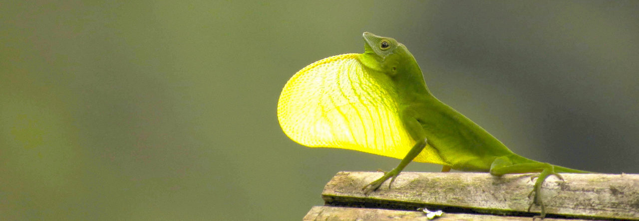
Anolis cybotes, one of the species included in Husak and Lovern, still showing its dewlap during copulation.

Anolis cybotes, one of the species included in Husak and Lovern’s study, still showing its dewlap during copulation.

If I were to take survey of Anole Annals readers regarding the factors that regulate aggressive and showy behaviors, I suspect that the vast majority of you would implicate testosterone as the primary culprit. Whether we are discussing humans or nearly any other vertebrate, there is a common societal notion that testosterone fuels these behaviors like oxygen fuels fire. The widespread belief is simple: individuals with more testosterone tend to exhibit more aggressive, ostentatious, and risky behaviors.

For decades researchers have investigated the link between testosterone and behavior in a variety of biological contexts – including different behaviors, experimental manipulations, environmental conditions, and life history parameters – but rarely in wild animals or within an evolutionary context. If the supposed testosterone-behavior correlation is extended to a broader, comparative context, it would suggest that aggressive species should also have higher levels of circulating testosterone than more placid species. But, in an upcoming paper, Husak and Lovern test the testosterone-behavior supposition among Anolis lizards and, quite frankly, turn it right on its head. To give away their conclusion at the outset, three of the four “aggressive” anole lineages examined have evolved this behavior without a clear correlation with circulating levels of testosterone.

Anolis lizards are renowned for their convergent anatomical evolution (reviewed in Lizards in an Evolutionary Tree), but these species have also independently evolved similar behaviors. In a study that was one of the first of its kind, Johnson et al. showed that the Anolis ecomorphs exhibit evolutionary convergence towards similar patterns of aggressive display and territorial behaviors. Trunk-ground anoles tended to be the most “aggressive” ecomorphs, consistently exhibiting higher display rates and  territoriality than the trunk-crown, grass-bush, or twig ecomorphs. Twig species tended to exhibit the least aggressive behavior in the analysis. (Also see Ord et al. 2013 for a more fine-scale dissection of display behavior.) Using this pattern of convergent behavior as a foundation, Husak and Lovern predicted that trunk-ground anoles would have higher levels of circulating testosterone than other ecomorphs from the same island, twig anoles the least. The absolute levels of testosterone might vary depending on the specific lineage in question, but they predicted that the rank-order of testosterone on each island would follow the behavioral continuum described in Johnson et al. In total the authors surveyed circulating levels of testosterone and corticosterone, an adrenal steroid hormone associated with stress, in 18 Anolis species!

Figure 1 from Husak and Lovern 2014: Circulating testosterone levels in 18 species of Caribbean Anolis lizards. Bars group by ecomorph classification (CG= crown giant, GB= grass-bush, T= trunk, TC = trunk-crown, TG = trunk-ground, TW= twig) and color coded by island (white = Bahamas, light gray = Jamaica, dark gray = Dominican Republic, black = Puerto Rico).

Figure 1 from Husak and Lovern 2014: Circulating testosterone levels in 18 species of Caribbean Anolis lizards. Bars group by ecomorph classification (CG= crown giant, GB= grass-bush, T= trunk,
TC = trunk-crown, TG = trunk-ground, TW= twig) and color coded by island (white = Bahamas, light gray = Jamaica, dark gray = Dominican Republic, black = Puerto Rico).

As I already stated, the authors found no support for the idea that elevated levels of circulating testosterone consistently drive aggressive behavior in Anolis lizards. Instead they found that three out of the four clades of trunk-ground anoles had the lowest levels of testosterone, the opposite pattern than would be predicted based on their behavior.

Transgenerational Effects Of Nutrition Observed In Anolis sagrei

Mothers affect the quality of their offspring. As humans, this seems obvious. For example, expecting mothers often take prenatal vitamins, limit their consumption of certain foods, and avoid kitty litter knowing that these minor environmental factors can affect the normal development of the fetus. Related statements could be made for the relationship of a mother and child after birth. Understanding the precise effects that parents have on their offspring has been of great interest to biologists from many disciplines as they disentangle the genetic and environmental factors that underlie differential survival and reproduction for individuals within a population (fitness). Because Anolis lizards can be easily maintained in captivity and their eggs readily manipulated they provide a useful model for the examination of maternal effects. Warner and Lovern took advantage of these qualities and tested the role of maternal body condition on offspring quality in the brown anole, Anolis sagrei.

A. sagrei from Cayman Brac

A. sagrei from Cayman Brac

Nutritional stress is a well-studied example of how maternal condition may affect juvenile quality; if the mother is malnourished the quality of her egg yolk may suffer, which, in turn, affects embryonic development. The authors tested this hypothesis in A. sagrei by manipulating the amount of food gravid females received, feeding approximately 168 crickets per lizard in a “high-prey” treatment versus 84 crickets in a “low-prey” treatment distributed over 11 weeks. During this time the authors carefully assessed the number and size (mass) of the eggs and, subsequently, the quality (mass-and-snout to vent length) of the hatchlings. Impressively, the authors didn’t stop there. They also experimentally manipulated the amount of nutrition in a subset of eggs by removing yolk with a syringe. Followed by a battery of statistical models, this study is quite a nice physiological analysis that has evolutionary implications.

When comparing the two diet regimes, Warner and Lovern found that body condition does affect the quality of offspring; females maintained on the “low-prey” diet produced eggs 6.6% smaller than females raised on the “high-prey” diet. In turn, smaller eggs also tended to hatch more quickly and smaller eggs produced smaller hatchlings, both probably due to the lower amount of available nutrition (paradoxically, neither incubation time or hatchling mass was directly correlated with maternal prey availability). Low prey availability also results in hatchlings with slower growth rates. The experimental reduction of egg yolk supports the results of the prey availability study: hatchlings from yolk-reduced females were 8% shorter and 23% lighter and grew more slowly than those hatched from unmanipulated eggs. It is clear from their results that nutrition has an effect on hatchling quality well into life, after the obvious maternal effects have passed. There are a number of other interesting correlations (and statistical caveats) described within the text that may also be of interest to some readers.

Figure 5 from Warner and Lovern 2014.

Figure 5 from Warner and Lovern 2014.

What is becoming clear from studies like these is that environmental stressors can have lasting effects on organismal development that transcend generational boundaries. Mechanistic studies, such as those on the American alligator, illustrate that these effects are mediated by heritable methylation patterns of key regulatory genes. The stressors do not need to be long lasting; physiological responses can result from acute events that occur within key developmental windows, often when a particular organ is maturing. While stressing the embryo too far results in abnormal embryonic development, more subtle effects may not arise until late in life or subsequent generations. Anoles, and A. sagrei in particular, may provide a number of opportunities for environmental health research in the future. Studies such as the one described above could be performed to more precisely dissect the organ-specific effects of maternal nutritional stress or whether the effects dissipate with age. Similar to the alligator studies, eggs laid in polluted soils may allow opportunities for developmental toxicology research. Growing genomic resources may allow for examination of genome-wide and gene-specific methylation patterns within and outside of polluted habitats. The possibilities are broad and the impact cannot be predicted at this time, but the potential is there for much more detailed mechanistic research on the relationship between developmental physiology and the environment.

Functional Genomics Comes to Anolis Lizards

One of the major experimental advances in recent decades has been the battery of methods capable of functionally validating hypotheses regarding the molecular networks that regulate biological processes. For biologists, these emerging methods allow us to move beyond descriptive and correlational studies to new dimensions where we can experimentally validate our observations. Until recently these technologies were, by and large, reserved for the most well developed laboratory model systems (e.g., mouse, chicken, zebrafish, Drosophila), systems that rarely have direct utility to ecologists and evolutionary biologists. But the topography of biology is changing. These methods are rapidly becoming more easily applied to non-model systems, such as our favorite genus Anolis. In an upcoming paper from the Menke Lab, the tools of functional genomics are applied to anole limb development, taking another step towards making Anolis a truly integrative model system.


In situ hybridization showing expression of of early limb genes in A. sagrei.

In situ hybridization showing expression of of early limb genes in A. sagrei.

Park et al. describe a micromass culture system to explore the molecular regulation of anole limb morphogenesis. In their protocol, Park et al. collect cells from early limb buds of A. sagrei, dissociate the cells from one another, and then add them to a dish as a small (i.e., micro) bolus (i.e., mass) of cells with the appropriate growth media. Even when removed from the embryo, these cells maintain the characteristics of limb cells, developing cartilage after about two weeks and maintaining their molecular signature for at least eight days. This small mass of cells can be grown for up to 30 days and, therefore, provide a useful template for experimental manipulations. More details of this protocol are described in great detail in the paper. Compared to other technologies which require far greater investment, their protocol should be accessible to anyone with access to a tissue-culture laboratory.

Anolis is an emerging model of limb development, but previous studies have focused on describing morphometric patterns of limb growth, not the molecular regulation of limb development. In fact, there have been no studies systematically dissecting the molecular regulation of limb development in any squamate species despite broad interest in this topic in the laboratory mouse and chick systems for 40 years. To study the molecular mechanisms regulating limb morphogenesis, Park et al. forced the expression of the gene Pitx1 – a hindlimb-specific molecule in mouse and chick – in micromass cultures derived from both forelimb and hindlimbs. This experiment verified that one step of the limb regulatory network, the relationship between Pitx1 and Hoxc11, is likely conserved among amniote lineages. While at this time this  may have been a proof of principal experiment, this protocol may have future implications for both developmental and evolutionary research in Anolis. For example, multiple transgenes can be readily cloned and incorporated into the micromass cultures. In addition, micromass cultures derived from species with distinct limb morphologies may also open to door to finding pathways that are regulated in novel ways across Anolis lizards.

Park et al. Figure 4

Binding domains of Pitx1 in the intergenic region of Hoxc11. Note conservation of binding region throughout mammals (shaded arrows), but lack of conservation among amniotes (white arrows).

Salmonella Found In Green Anoles

Sorry to post this right before Thanksgiving dinner, but here is something to think about the next time you eat after handling an anole: approximately 30% of anoles in Japan carry salmonella, twice the level in feral goats and more than ten times greater than that found in public toilets. Read the complete (freely available) study here.

Salmonella in green anoles


Where Do Lizards Go When It’s Cold?

When I arrived at the University of Florida this summer, I was struck by the bustling sidewalks – bustling not with students, but with brown/festive/Cuban anoles. They were everywhere! But now that it’s cool out (not cold, lets say below 70 degrees to be generous), they are essentially gone. Where I could once count ten anoles sitting on a single bench, it would now take some effort to find this many in a reasonable amount of time.  Where did they go?

Carrig and his cold lizardsToday, while cleaning up piles of leaves from the yard, I was surprised to find a fairly large collection of A. sagrei under my leaf piles. One pile had as many as four under it (lizards that were not shoveled up inadvertently before realizing my cache) and every pile had at least a few. These individuals were almost certainly below their thermal optima as my son had no problem scooping up three at once and proudly displaying them for a photo op. Is it possible that the lizards found these piles warmer than those found elsewhere, attracted to the heat of the composting leaves? Maybe they were just there to stay out of yesterday’s rain and had not yet ventured out. I would be curious to hear if anyone else ever observed something like this?

The Evolution Of Squamate Developmental Sequences

A. sagrei developmental sequence. Cover illustration from Sanger et al. (2012) Proc. B.

A. sagrei developmental sequence. Cover illustration from Sanger et al. (2012) Proc. B.

As discussed previously in the Annals, interest in squamate development is rapidly accelerating. Our growing community makes this an exciting time to study lizard development, especially in a comparative context. A recent study by Andrews et al. capitalizes on the increasing number of developmental resources for squamates to assess variability in developmental sequences across lizards and snakes. One of our favorite anoles, Anolis sagrei, represents one of the 21 species included in this study. The conclusions of this study speak to several long-standing evolutionary questions and opens up new avenues of investigation that may be of interest the readers of this blog. 


A $90 Night Light?

Several years ago I reviewed some lighting options for people interested in hunting anoles at night (Who wouldn’t? Throw that noose away!). At the time I recommended several readily available lights ranging from 300 to 700 lumens, $60 to $500 respectively. Needless to say, most anole enthusiasts were likely priced out of the brightest lights. However, I recently found an option affordable to even our dedicated summer field assistants. A relatively new company to the US, Magicshine, advertises an 1100 lumen light for only $90, the MJ-808U.

Maginshine MJ 808U

Maginshine MJ 808U

Now the first thing we should all do is assume that this is too good to be true. In just a few years our discussion has gone from $500 to $90 for a supposedly superior light. Come on! Online reviews of Magicshine’s products are generally favorable, but mixed. Reviews on mountain bike forums comment on the relatively poor construction and historically bad batteries. Several reviewers have also commented on overheating problems for riders not peddling their hardest.The best part of Magicshine is by far their price. But the last I checked we rarely look for anoles on bikes at night so we will need to take all of this in with some hesitation.

So what about herping? Time will ultimately tell how these lights hold up to our uses. I received my light in the mail earlier this week and have used it twice. To keep my hands free I also purchased the accessory head strap for about $8. I also need to purchase the extension cable as the attached cable between the battery and light is too short to reach my backpack from my head. First impressions, the light is retinal burning bright. I have absolutely no complaints there. I will warn you now, however, that the light gets hot, but has yet to overheat for me. I haven’t fully run down the battery yet either, but it has lasted for over 90 minutes of burn time so far. In summary, my first impressions are as the online reviews suggests, there are both pros and cons to this product, but for $90 why not give it a try. I am cautiously optimistic.

Has anyone else discovered this light yet? If so, please share your reviews with the community.


Anolis Chlorocyanus In Florida?

Years ago I heard reports that Anolis cholocyanus had established a small population in  Dade County. Does anyone know if this population is still viable? I am starting a new project and it would be quite beneficial to examine a few living specimens before investing in a full trip to the Dominican Republic. If anyone has any information about the status of this introduced species I would be extremely grateful to learn more.

Have a good weekend!


Page 3 of 6

Powered by WordPress & Theme by Anders Norén