Author: Simon Baeckens

Evolutionary ecologist and functional morphologist interested in what drives and constrains phenotypic diversity in lizards, and how it relates to organismal function.

Repeated Evolution of Skin Surface Micro-Architecture and Increased Hydrophobicity in Semi-Aquatic Anoles

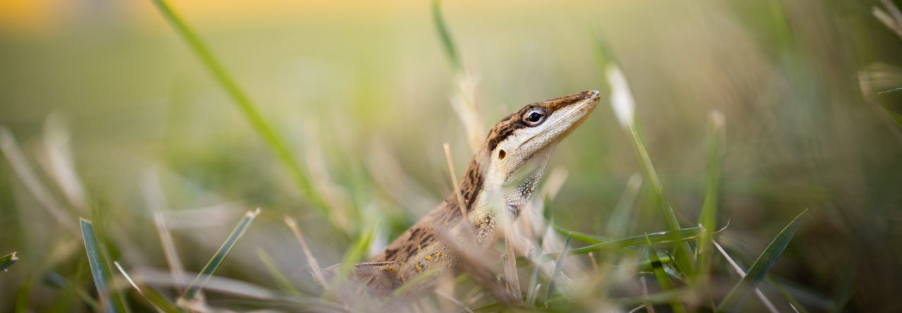
A Stream Anole (Anolis oxylophus) on the cover of the Journal of Experimental Biology (vol. 224, issue 19). Photo credit: © Day’s Edge Productions.

Terrestrial animals that venture into the water on a regular basis face a number of challenges not encountered by their strictly terrestrial counterparts. While submerged, they must deal with hydrodynamic drag forces hindering locomotion and with the risk of running out of air. Back on land, the film of water adhering to their body surface may interfere with locomotion and thermoregulation or may increase the risk of bio-fouling. Many semi-aquatic invertebrates (and plants) have developed complex surface microstructures with water-repellent properties to overcome these problems, but equivalent adaptations of the skin have not been reported for vertebrates that encounter similar environmental challenges.

The transition to a semi-aquatic lifestyle has independently occurred multiple times throughout the evolutionary history of Anolis (see Fig. 1A below). In anoles, the skin surface is covered with microscopic hair-like ornaments, and contingent upon its complexity, organization, and length dimensions, these hair-like microstructures may have the potential to generate extreme surface hydrophobicity. Indeed, similar skin surface microstructures have been found in geckos and are shown to be responsible for the highly hydrophobic surface of their skin. The water-resistant properties of anole skin, however, have remained unexamined, but very recent discoveries have provided valuable insight into this matter. Boccia et al. (2021) observed that semi-aquatic Anolis lizards are able to sustain long periods submerged underwater by iteratively expiring and re-inspiring narial air bubbles. As in semi-aquatic insects, a hydrophobic skin is a key requirement for the underwater formation of an air bubble, hence, functional respiration, so a hydrophobic skin in semi-aquatic anoles is implied. However, whether a hydrophobic structured skin surface in anoles has evolved in response to life at the water-land interface is still an open question. Answering this question was the primary goal of our study.

We studied the skin surface morphology of preserved anole specimens using scanning electron microscopy and tested the wettability of the skin surface using contact-angle goniometry (Fig. 1D). We found that the skin surface of semi-aquatic species of Anolis lizards is characterized by a more elaborate microstructural architecture (i.e. longer hair-like structures; Fig 1B,C) and a lower wettability (Fig. 1D,E) relative to closely related terrestrial species. In addition, phylogenetic comparative models revealed repeated independent evolution of enhanced skin hydrophobicity associated with the transition to a semi-aquatic lifestyle, providing evidence of adaptation.

Figure 1 from Baeckens et al. (2021)

We believe our findings bring an additional dimension to the recent biological phenomenon described by Boccia et al. (2021) namely that diving Anolis lizards not only repeatedly and independently evolved a specialized rebreathing behavior with the transitioning to a semi-aquatic lifestyle, but that its evolution presumably also coincided with, or was preceded by, the evolution of a hydrophobic structured skin to successfully do so.

Our study was published in Journal of Experimental Biology. And thanks to Day’s Edge Productions, we also got the issue cover!

Reference

S. Baeckens, M. Temmerman, S. Gorb, C. Neto, M. Whiting & R. Van Damme (2021) Convergent evolution of skin surface microarchitecture and increased skin hydrophobicity in semi-aquatic anole lizards. Journal of Experimental Biology 224(19): jeb242939 (doi: 10.1242/jeb.242939)

Visualising the 3D Surface Structure of Lizard Skin

Imaging the surface topography of lizard skin using gel-based stereo-profilometry.
(Baeckens et al. 2019)

The skin surface structure of lizards varies greatly among species, likely because it plays a key role in a range of tasks, such as camouflage, locomotion, self-cleaning, mitigation of water loss and protection from physical damage. Yet, we still know remarkably little about how variation in skin surface structure translates to functional variation. Part of this gap in our understanding can be traced back to the lack of means to perform high-throughput and detailed analysis of the 3D anatomy of lizard skin in a non-destructive manner.

To tackle this hiatus, I was fortunate enough to be able to round up a great team of scientists and to start exploring the possibilities of a new imaging technique, termed gel-based stereo-profilometry. In this approach, a deformable transparent gel pad with one opaque surface is pressed onto the object of interest, creating a surface impression. While the gel pad is still in contact with the object, a series of photographs from six different illumination angles are acquired, and a topographical 3D map of the surface is created by merging the acquired images using
specialized surface analysis software.

Using this technique, we successfully imaged the 3D skin surface structure of Anolis cristatellus specimens in great detail (pixel resolution of 0.86 µm) and in a short-time frame (average acquisition time of imaging and digital reconstruction combined was 90 seconds). In our new paper, we demonstrate that this technique is exceptionally useful for the rapid 3D structural characterisation of lizard skin surfaces without any specimen preparation, permitting 3D visualization in situ and even in vivo. This technique opens exciting new avenues for investigating structure–function relationships in lizard skin.

In addition to the ability to quantify the micro- and macro-structural details of lizard skin, the 3D data sets acquired using gel-based stereo-profilometry can be directly converted into surface meshes, which can in turn be 3D printed. These tangible models can then be directly employed for studies to investigate the role of scale geometry on animal–substrate interactions, or enlarged for educational purposes to illustrate key differences between different squamate taxa.

3D print of lizard skin.

Baeckens S, Wainwright DK, Weaver JC, Irschick DJ & Losos JB (2019) Ontogenetic scaling patterns of lizard skin surface structure as revealed by gel‐based stereo‐profilometry. Journal of Anatomy 235, 346–356.

Drivers and Constraints of Within-Species Diversity in Dewlap Design

Sampling locations of the populations of study across the Caribbean. (1) Soroa (Cuba), population 1; (2) Soroa (Cuba) population 2; (3) Grand Cayman; (4) Santa Clara (Cuba); (5) South Bimini; (6) Chub Cay; (7) Andros; (8) Crooked Island; (9) Acklins; (10) San Salvador; (11) Staniel Cay; (12) Pidgeon Cay; (13) Grand Bahama; (14) South Abaco; (15) Cayman Brac; (16) Little Cayman; (17) Jamaica.

The dewlap is arguably one of most fascinating features of anoles. For me, it is the baffling diversity in dewlap size, coloration, and use —both among and within species— that makes it so interesting. However, understanding the origin and evolution of dewlap diversity in Anolis has proven a daunting task (Nicholson et al. 2007; Vanhooydonck et al. 2009). In an attempt to make (a little more) sense of the drivers and constraints of anole dewlap variation, a team of Belgian researchers from the University of Antwerp, led by evolutionary ecologist Tess Driessens, decided to look at dewlap diversity in Anolis sagrei. They surveyed 17 island populations of A. sagrei across the Caribbean and quantified dewlap design (color, size) and dewlap display behavior of both males and females.

Last year, Driessens and colleagues published their findings on how variation in abiotic factors (such as precipitation, temperature and other climatic variables) could explain much of the observed inter-island variation in dewlap design and use in A. sagrei (‘signal efficacy’ hypothesis). In a paper that came out last week, the team reports on the role of the biotic environment in driving dewlap diversity in the brown anole. Inspired by the wonderful study of Vanhooydonck et al. (2009), the researchers tested whether among-population dewlap variation could be (at least partially) assigned to variation in predation pressure (estimated by island size, tail break frequency, presence/absence of the predatory curly-tailed lizards, clay model attack rate), sexual selection (using sexual size dimorphism), and/or species recognition (number of syntopic Anolis species). Overall, they found only limited support for the idea that the extensive interpopulational variability in dewlap design and use in A. sagrei is mediated by variation in their biotic environment. Although they did find that males from larger islands show higher dewlap display intensities than males from smaller islands, and that males are more likely to have a ‘spotted’ dewlap pattern when co-occurring with a high number of syntopic Anolis species, the direct connection with predation pressure and species recognition remains ambiguous and demands further investigation.

In another recent paper, focusing only on the size of the male dewlap and their maximum bite capacity, the Belgian researchers asked a different question: does dewlap size signal fighting capacity (estimated by bite force) in A. sagrei, and is this true for all 17 sampled populations? And, does the level of signal honesty (that is, the steepness of the dewlap size-bite force relationship within a population) vary among populations, and is it linked with the strength of intrasexual selection? Their results showed that absolute dewlap size is an excellent predictor of bite force in all A. sagrei populations. However, relative dewlap size was only an honest signal of bite performance in 4 out of the 17 populations. Surprisingly, the level of signal honesty did not correlate with the strength of intrasexual selection.

Male brown anole biting on a purpose-built force plate. Photo by Tess Driessens

While the work of Tess Driessens and her team sheds new light on the drivers and constraints of dewlap diversity in A. sagrei, there is still plenty of study material left for future dewlap fanatics.

Evidence for evolutionary determinism in the signal design of lizards?

Photographs of a subset of lacertid lizard species used in this study. From the left top to the right bottom: Acanthodactylus beershebensis, Lacerta bilineata, Dalmatolacerta oxycephala, Podarcis melisellensis, Tropidosaura gularis, Podarcis siculus, Heliobolus lugubris, Algyroides nigropunctatus, Lacerta media.

Photographs of a subset of lacertid lizard species used in this study. From the left top to the right bottom: Acanthodactylus beershebensis, Lacerta bilineata, Dalmatolacerta oxycephala, Podarcis melisellensis, Tropidosaura gularis, Podarcis siculus, Heliobolus lugubris, Algyroides nigropunctatus, Lacerta media.

The vast array of signals used in animal communication is a continuous source of awe and a hot topic in evolutionary and behavioral research. One important factor contributing to the signal diversity we witness today is ‘signal efficacy’: the ability of a signal to travel efficiently through the environment and attract the receiver’s attention. With this in mind, natural selection is expected to mold signal design for maximum efficacy of information transmission and detectability, leading to signal variation among populations/species living in different environments. To illustrate, a recent study by Tess Driessens and colleagues assessed the degree of variation in the dewlap design of Anolis sagrei by comparing 17 populations distributed across the Caribbean (Fig. 1).

Phylogenetic relationships among seventeen Anolis sagrei populations. Pie charts illustrate dewlap pattern proportions for each population per sex (black = solid; light grey = marginal; dark grey = spotted). Photographs represent male and female dewlaps of typical individuals from every population.

Fig. 1 — Phylogenetic relationships among 17 Anolis sagrei populations. Pie charts illustrate dewlap pattern proportions for each population per sex (black = solid; light grey = marginal; dark grey = spotted). Photographs represent male and female dewlaps of typical individuals from every population.

Their findings showed large interpopulational variation in dewlap size, pattern, and color, and more interesting, they established a link between the dewlap design of brown anoles and the environment they live in. Lizards occurring in more ‘xeric’ environments had a higher proportion of solid dewlaps with a higher UV reflectance; lizards inhabiting ‘mesic’ environments had predominantly marginal dewlaps showing high reflectance in red. This was true for both males and females. Like Ng et al. (2011) and their observations on dewlap variation in A. distichus across an environmental gradient, Driessens et al. (2017) interpret their findings as evidence for adaptive divergence of a signaling apparatus.

Surprisingly though, while there are numerous great examples of comparative studies finding support for convergent evolution in visual and acoustic signaling systems, (e.g. Endler 1992; Fleishman 1992; Nicholls & Goldizen 2006, to say a few), similar (comparative) studies, but then, on the phenotype of chemical signals are almost entirely lacking. This is probably due to the combination of only very recent developments in chemical analytical and statistical comparative tools, the time researchers need to assemble a large-scale multi-species chemical dataset, and perhaps due to our own predisposition to visual and auditory signals. Currently, the proper analytical tools for studying natural products chemistry are available and affordable, permitting comprehensive taxon-wide research on the evolution of chemical signal diversity and design. Ultimately, there has never been a better time as now to be a comparative chemical ecologist.

Photograph of the cloacal region of a male lacertid lizard (Lacerta agilis), showing his numerous femoral pores with protruding glandular secretion.

Photograph of the cloacal region of a male lacertid lizard (Lacerta agilis), showing his numerous femoral pores with protruding glandular secretion.

Finally, three Belgians, two Spaniards and one Greek (sounds like the start of a joke with ample potential) took up the challenge to examine variation in the chemical signal design of lizards. Although underrepresented in studies on chemical signal diversity, lizards are an excellent group for investigating chemical signal evolution, as many of them they bear numerous glands on their thighs that secrete waxy substances, which they deposit while moving through their habitat. These secretions are often considered the leading source of chemical signals involved in lizard communication.

The study started with a quest. A quest to collect gland secretions of as many species as possible (within a PhD timeframe). Luckily, we were fortunate enough to be able to count on the help of many collaborators (Shai Meiri, Chris Broeckhoven, …). We focussed on lacertid lizards, as they are a species-rich family distributed over a wide geographical area, and known to rely strongly on chemical communication in several contexts.

In total, we sampled secretions from 64 species throughout, Europe, Africa, and Asia, covering a wide array of habitats and climate regions: from the Mediterranean maquis over the alpine meadows in the Pyrenees Mountains, to the sandy Israeli dunes and the Kalahari Desert of South Africa (Fig. 2). Back in the lab, we determined the chemical composition and chemical ‘richness’ (number of different chemical compounds) of the secretions using GC-MS, and obtained climate data for all catch-localities from online databases.

Map showing the sample localities of the 64 lizard species under study.

Fig. 2 — Map showing the sample localities of the 64 lizard species under study.

Our gathered data showed considerable variation in the chemical richness and composition of lacertid secretion. Shared-ancestry failed to explain among-species patterns of variation, hinting that chemical signals may change relative rapidly. Most interestingly, our findings revealed a strong relationship between the environmental conditions species live in and the chemical composition of their glandular secretions. On the one hand, lizards living in ‘xeric’ environments, characterized by high temperatures and arid conditions, contained higher proportions of stable and heavy-weight compounds in their secretions. Hot and dry conditions increase the evaporation rate of chemicals, and so, decreasing the longevity of a signal. Stable and heavy-weight compounds most likely reduce evaporation rate and counteract the rapid signal fade-out through evaporation, generating a highly persistent scent-mark. On the other hand, species inhabiting wet, humid conditions produced highly aromatic and low-weight secretions containing numerous different compounds. This chemical mix probably creates a volatile-rich signal that can be used for long-distance airborne communication.

While we cannot deny that these findings of convergent evolution in the design of chemicals signals are fascinating, some would say this outcome is not unexpected.

“[…] a cadre of scientists has taken the […] view, that convergence is the expectation, that it is pervasive, and that we should not be surprised to discover that multiple species […] have evolved the same features to adapt to similar environmental circumstances. From this perceived ubiquity, the scientists draw a broader conclusion: evolution is deterministic, driven by natural selection to repeatedly evolve the same adaptive solutions to problems posed buy the environment. — J. Losos (Improbable Destinies, p. 33)

Nonetheless, I am confident to state that using by far the largest comparative dataset amassed to-date to examine patterns of chemical signal divergence, we have provided strong evidence for a significant relationship between chemical signal design and prevailing environmental conditions, which may results from differential selection on signaling efficacy (Baeckens et al. 2017).

Powered by WordPress & Theme by Anders Norén