Twenty exquisitely preserved anole fossils in 20 My old Dominican Amber have been reported on in a paper out in Proceedings of the National Academy of Sciences (PNAS) this week.
Previously on AA, I reported that the search was on to find anole fossils in order to piece together the anole family tree. We were extremely fortunate to find in the end 38 amber fossils with anole inclusions, sourced from museums such as the Staatliches Museum für Naturkunde Stuttgart, Germany, American Museum of Natural History, and Naturhistorisches Museum, Basel Switzerland, as well as from generous private collectors.
All of the fossils were exquisite, stunningly-preserved anoles in Dominican Amber. Sometimes just a foot or tail was preserved, sometimes a whole limb or two, or an isolated head, but occasionally a whole lizard was preserved laid out as if it has been pressed into resin just moments before.

Modified from Figure 1 of Sherratt et al. 2015 PNAS.
Using micro-CT scanning to peer inside the fossils, we were delighted to find well-preserved skulls and skeletons. We were surprised to find that many of the amber pieces had air-filled pockets representing where the lizard body had once been (but subsequently mostly rotted away), and the scales had left their impression on the amber. This allowed us to view the scales of the limbs and toepads in the greatest of detail.

The forelimb lying atop belly scales of a trunk-ground fossil, specimen M of Sherratt et al. 2015.
Twenty of these fossils were complete enough, or preserved with the right body parts (limbs with a pelvis, or toepads with countable lamellar scales) to study qualitatively. I micro-CT scanned 100 modern specimens from the Harvard MCZ collection, representing adults and juveniles of all the ecomorphs in Hispaniola. With these data, I build up a dataset of measurements of the limbs, skulls and pelvic girdles that could be used to compare with the fossils. Working fossil by fossil, I used discriminant function analysis to assess the probability that the fossil matched each of the modern ecomorphs.

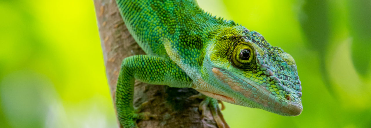
The fossil twig anole, from Jose Calbeto of Puerto Rico.
The results were very exciting. We found evidence for four of the six ecomorphs in the amber. Trunk-crown were the most abundant, but there was also one that fell within the twig anoles, two that fell with trunk and two with trunk-ground anoles. Not all the fossils could be assigned to an ecomorph with high probability. Though, my gut feeling is that there is a second twig anole (specimen P) based on the distinct few lamellar scales on its widely-expanded toepads, but sadly it didn’t have enough skeleton and no hind limbs preserved to add to the analysis.
We didn’t find any fossils that resembled crown-giants or grass-bush anoles. Why?


