
(Baeckens et al. 2019)
The skin surface structure of lizards varies greatly among species, likely because it plays a key role in a range of tasks, such as camouflage, locomotion, self-cleaning, mitigation of water loss and protection from physical damage. Yet, we still know remarkably little about how variation in skin surface structure translates to functional variation. Part of this gap in our understanding can be traced back to the lack of means to perform high-throughput and detailed analysis of the 3D anatomy of lizard skin in a non-destructive manner.
To tackle this hiatus, I was fortunate enough to be able to round up a great team of scientists and to start exploring the possibilities of a new imaging technique, termed gel-based stereo-profilometry. In this approach, a deformable transparent gel pad with one opaque surface is pressed onto the object of interest, creating a surface impression. While the gel pad is still in contact with the object, a series of photographs from six different illumination angles are acquired, and a topographical 3D map of the surface is created by merging the acquired images using
specialized surface analysis software.
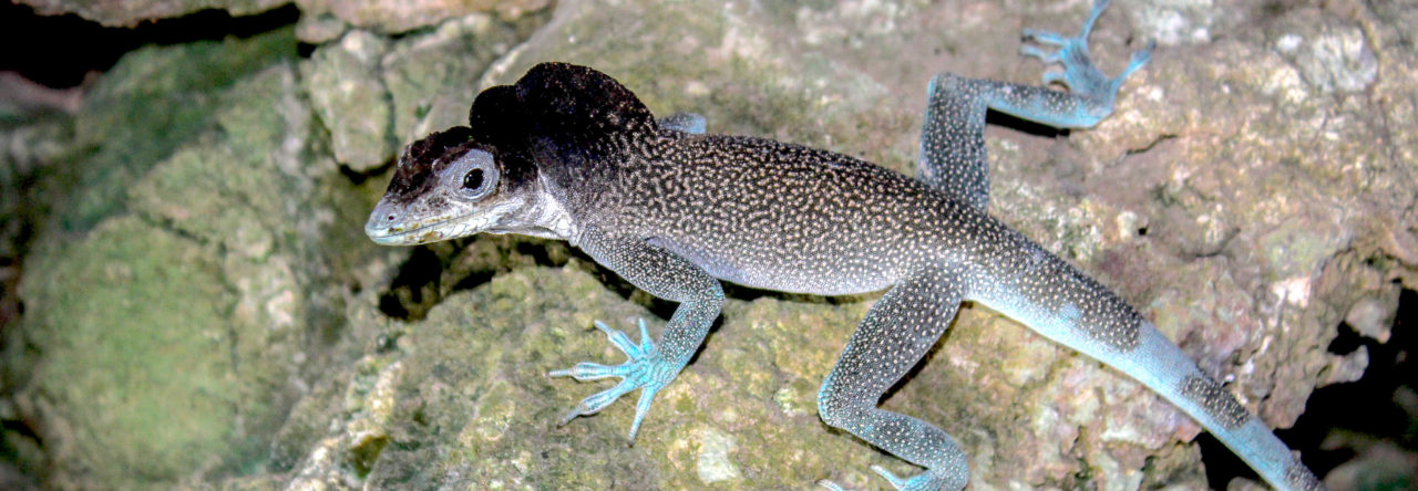
Using this technique, we successfully imaged the 3D skin surface structure of Anolis cristatellus specimens in great detail (pixel resolution of 0.86 µm) and in a short-time frame (average acquisition time of imaging and digital reconstruction combined was 90 seconds). In our new paper, we demonstrate that this technique is exceptionally useful for the rapid 3D structural characterisation of lizard skin surfaces without any specimen preparation, permitting 3D visualization in situ and even in vivo. This technique opens exciting new avenues for investigating structure–function relationships in lizard skin.
In addition to the ability to quantify the micro- and macro-structural details of lizard skin, the 3D data sets acquired using gel-based stereo-profilometry can be directly converted into surface meshes, which can in turn be 3D printed. These tangible models can then be directly employed for studies to investigate the role of scale geometry on animal–substrate interactions, or enlarged for educational purposes to illustrate key differences between different squamate taxa.

Baeckens S, Wainwright DK, Weaver JC, Irschick DJ & Losos JB (2019) Ontogenetic scaling patterns of lizard skin surface structure as revealed by gel‐based stereo‐profilometry. Journal of Anatomy 235, 346–356.

Leave a Reply