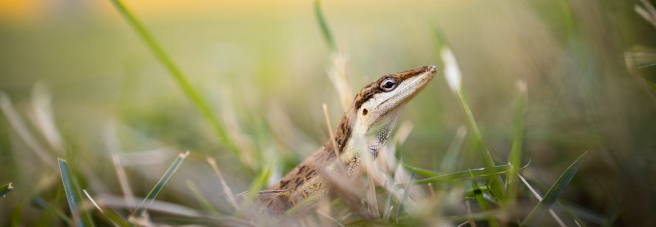
Back in September, we saw an Anolis carolinensis with a bizarre skeletal anomaly, the zig-zag tail. Several readers commented that this was quite a common trait, especially among captive lizards. I wanted to continue this theme with a curious Anolis cybotes specimen I found while CT scanning.
This image of a volume rendering of the skeleton shows a typical A. cybotes male pelvis, where the ilia articulates with the sacral vertebrae (denoted by arrow).
Now, the image below shows R186747, a male A. cybotes collected by Luke Mahler in the Dominican Republic. The lateral process of the first tail vertebrae has been adopted to form the sacrum on the left side, while the right remains standard, and the right side of the pubis appears to have an old healed fracture.
When I discussed this with my colleague, Kevin Mulder, he immediately brought up the following x-ray images of A. smaragdinus and A. sagrei (left to right) specimens (and he has several others) that also have asymmetrical sacral vertebrae and deformed transverse processes on the tail vertebrae. Therefore it appears this condition may be more common…
My gut feeling for the A. cybotes specimen was that this is more likely due to post-hatching trauma (such as escaped predation incident), rather than a developmental abnormality, but now I am not so sure. Do any readers have any ideas?
Regardless, it appears not to be a problematic condition since all of these animals were seemingly growing and living happily prior to being noosed!
- 20-Million-Year-Old Fossils Reveal Ecomorph Diversity in Hispaniola - July 27, 2015
- Anolis proboscis: Ugly and Famous - September 5, 2014
- The Fossil Species Anolis electrum Gets an X-ray Makeover - August 14, 2014


Hastley
As I understand it, similar vertebral anomalies are widespread in snakes, usually due to temperature fluctuations during incubation/gestation. In snakes, because the ventral scales have a 1:1 correspondence with vertebrae, you can actually check externally – aberrant vertebrae will show up as forked or incomplete ventrals. I can try to dig up the ref if you like.
Dmenke
Variation in vertebrae morphology has been observed in mice at the transition point from lumbar to sacral vertebrae. There appear to be both genetic and environmental influences that contribute to this variation, and some individuals exhibit an asymmetric vertebra at the lumbar/sacral transition with a more lumbar-like morphology on one side and a sacral-like morphology on the other.
http://dev.biologists.org/content/2/2/149.full.pdf
http://dev.biologists.org/content/21/1/97.full.pdf
Rich Glor
It would be interesting to start cataloguing these and other skeletal deformities in anoles. Can your CT scanner handle hatchling anoles? We occasionally get deformed hatchlings from our breeding experiments and it would be interesting to know if there is any consistency to the types of skeletal problems they’re emerging with.
Em Sherratt
Certainly the size of the hatchlings wouldn’t be a problem, but the degree of ossification may. But there are stains for cartilage that can be used on fresh material so that there is significant difference in x-ray attenuation between the materials. So yes, possible.
scantlebury
Yes, I have had numerous gecko species hatch over the years with kinked tails. Generally speaking, these deformities were associated with the incubator getting excessively hot, or the eggs showing mold growth (which itself is usual associated with bad incubation conditions).
Yoel Stuart
Chad Watkins from UT Arlington has been studying this very phenomenon of the vertebral-process being fused to the sacrum and its relation to HOX genes. He presented at the Evolution meetings (http://anoleannals.wordpress.com/2011/06/21/evolution-meeting-2011-homeotic-mutations-in-anoles/) and is currently analyzing hundreds of x-rays in addition to the genetics of carolinensis and sagrei, among other species.
Emma Sherratt
As a follow up on this, I was reading Hoffstetter and Gasc’s chapter on Vertebrae and Ribs in Biology of the Reptilia Vol 1 (1969), and there on page 264 is a drawing of exactly this condition (described from the lizard Gerrhosaurus flavigularis). So a well-known phenomenon, yet that doesn’t take away from the shear oddity of this condition.